Mice and tissue collection
C57BL/6J129Sv mice were used in this study. To collect timed-pregnant tissues, breeding pairs were mated in the morning, and vaginal plugs were checked in the afternoon. The presence of a vaginal plug was designated as day 0 of pregnancy. Pregnant uteri were collected from gestational days 3 to 9. Implantation sites were harvested and prepared as frozen tissue blocks by immersing them in OCT (Tissue-Tek, Thermo Fisher Scientific, Inc., Waltham, MA, USA) and solidifying them with liquid nitrogen vapor. The blocks were stored at −80 °C until processing. All animal experiments were conducted in accordance with the National Institutes of Health Guide for the Care and Use of Laboratory Animals humane animal care standards, and were approved by the Institutional Animal Care and Use Committee of the University of Vermont.
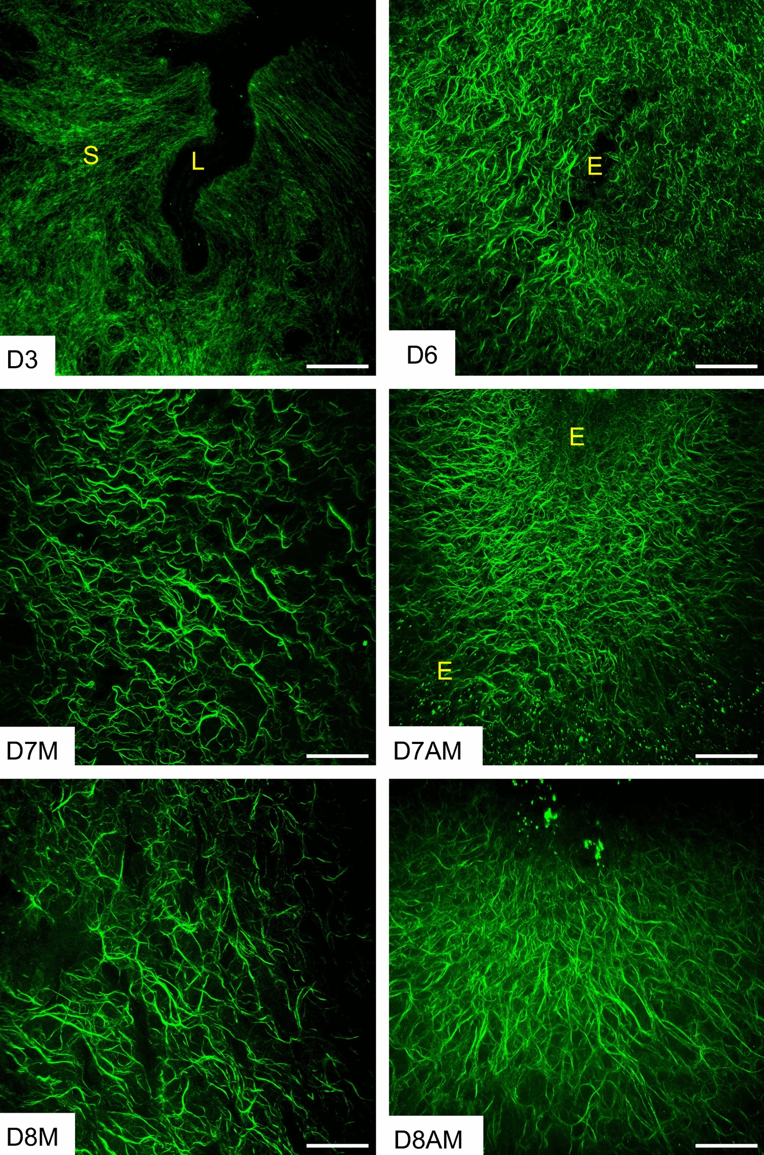
Second harmonic generation imaging
Frozen uterine cross sections of 50 μm thickness were utilized in this experiment. The thawed sections were subsequently covered with 0.1 M phosphate-buffered saline (PBS) to preserve the hydration of the tissue sections during imaging. A Zeiss LSM7 inverted microscope (Carl Zeiss AG, Oberkochen, Germany) equipped with an Achroplan 20x/0.8 objective lens was employed to acquire images of the slides. A Chameleon XR pulsed Ti: sapphire laser (Coherent, Santa Clara, CA, USA) was adjusted to emit a wavelength of 900 nm onto the tissue, resulting in a signal detected at 450 nm. The acquired images were subsequently analyzed using ImageJ software (National Institutes of Health).
Confocal microscopic imaging
Frozen uterine cross sections (5 µm thick) were fixed in acetone for 10 min and subsequently blocked with 10% normal goat serum (Life Technologies, Carlsbad, CA, USA) for 30 min at room temperature. Primary antibodies—COL1A1 (Cell Signaling Technology, Danvers, MA, USA; 72026, RRID:AB_2904565, 1:200), collagen 3 (Proteintech, Rosemont, IL, USA; 22734-1-AP, RRID:AB_2879158, 1:500), elastin (Elastin Products Company, Owensville, MO, USA; PR385, RRID:AB_3099530, 1:250), LOXL1 (Thermo Fisher Scientific, Inc., Waltham, MA, USA; PA5-87701, RRID:AB_2804357, 1:500), LOXL2 (Abcam, Waltham, MA, USA; Ab96233, RRID:AB_10677617, 1:100), LOXL3 (Thermo Fisher Scientific, Inc., Waltham, MA, USA; PA5-48462, RRID:AB_2633919, 1:500), LOXL4 (Thermo Fisher Scientific, Inc., Waltham, MA, USA; PA5-115520, RRID:AB_2900156, 1:100), and smooth muscle actin (Santa Cruz Biotechnology, Inc, Dallas TX, USA; sc-32251, RRID:AB_262054, 1:1000)—were diluted in blocking solution, added to the tissue sections. The slides were then incubated overnight at 4 °C. The following day, slides were washed with PBS and then incubated with secondary antibodies—anti-rabbit IgG Alexa Fluor 555 (Thermo Fisher Scientific Inc., Waltham, MA, USA; A32732, RRID: AB_2633281, 1:500) and anti-mouse IgG Alexa Fluor 488 (Thermo Fisher Scientific, Inc., Waltham, MA, USA; A11029, RRID: AB_2534088, 1:500)—in blocking solution for 30 min at room temperature. Slides were washed with PBS and mounted with ProLong Gold Antifade Mountant with DNA Stain DAPI (Thermo Fisher Scientific, Inc., Waltham, MA, USA). Images were acquired using a Nikon A1R confocal microscope (Nikon Instruments, Melville, NY, USA) equipped with a galvanometer scanner and illumination point scan at a rate of eight frames per second for a 1024 × 1024-pixel field. Both 4× and 10× objective lenses were used for imaging. Nikon NIS-Elements software (Nikon Instruments, Melville, NY) was used for image acquisition. The acquired images were further analyzed using ImageJ (National Institutes of Health).
Mouse endometrial stromal cell culture
On gestational day 3, the pregnant uteri were collected, sectioned longitudinally, and subsequently cut into 3–5 mm pieces. These tissues were subsequently digested with a mixture of 6 g/L dispase (07923, STEMCELL Technologies, Cambridge, MA, USA), 25 g/L pancreatin, and antibiotic–antimycotic solution ( Thermo Fisher Scientific, Inc., Waltham, MA, USA) in Hank's Balanced Salt Solution (HBSS) for 1 h at room temperature, followed by 15 min at 37 °C. After aspirating the enzyme mixture, the tissues were then digested with 250 µg/mL Liberase (Sigma-Aldrich, Saint Louis, MO, USA) in HBSS (Thermo Fisher Scientific, Inc., Waltham, MA, USA) for 45 min at 37 °C. The enzyme action was terminated by adding 10% fetal bovine serum (FBS; Thermo Fisher Scientific, Inc., Waltham, MA, USA). The mixture was filtered through a 70 µm strainer and subsequently centrifuged. The resulting pellet was diluted in DMEM-F12 (Thermo Fisher Scientific, Inc., Waltham, MA, USA) containing 2% FBS, 1% antibiotic and antimycotic solution, 1 µM progesterone (Sigma-Aldrich, Saint Louis, MO, USA), and 10 nM 17β-estradiol (Sigma-Aldrich, Saint Louis, MO, USA). Cells were counted using a hemocytometer and were plated in six-well plates at a concentration of 1 × 10⁶ cells/well. After 2–3 h, the culture medium was replaced, and thereafter the culture medium was replaced daily. Cell lysate and conditioned media were collected at 24, 48, 72, and 96 h for gene and protein expression analysis.
Quantitative polymerase chain reaction (qPCR)
Total RNA was extracted from stromal cell lysates using the RNeasy Mini Kit (74104, Qiagen, Germantown, MD, USA), following the manufacturer’s protocol. Complementary DNA (cDNA) was synthesized using the iScript Reverse Transcription Supermix (Bio-Rad Laboratories, Hercules, CA, USA). Quantitative PCR was performed using SYBR Green (Thermo Fisher Scientific, Inc., Waltham, MA, USA) and primers specifically designed for the genes of interest. Gene expression was quantified using the 2–ΔΔCt method, with target gene expression normalized to the housekeeping gene Rplp0. The primers used in this study are listed in Table 1.
Table 1 The list of primers used in this studyWestern blot
Endometrial stromal cells were lysed using radioimmunoprecipitation assay (RIPA) buffer containing 1% protease and phosphatase inhibitors (Thermo Fisher Scientific Inc., Waltham, MA, USA) and stored at −80 °C until processing. Protein concentration was determined using a bicinchoninic acid (BCA) protein assay (Thermo Fisher Scientific, Inc., Waltham, MA, USA). Samples (20 µg) were boiled at 95 °C for 10 min in Laemmli Sample Buffer (Bio-Rad Laboratories, Hercules, CA, USA) with β-mercaptoethanol (Sigma-Aldrich, Saint Louis, MO, USA). The samples and a protein standard (Precision Plus Protein Kaleidoscope, Bio-Rad Laboratories, Hercules, CA, USA) were loaded into a 10% Bis/Tris–HCl sodium dodecyl sulfate–polyacrylamide gel electrophoresis (SDS-PAGE) gel and separated at 50 V for 10 min, followed by 100 V for 1 h. Proteins were then transferred onto a nitrocellulose membrane (Bio-Rad Laboratories, Hercules, CA, USA) at 100 V for 1 h at 4 °C. Membranes were blocked with 3% blotting-grade nonfat dry milk in TBST (Bio-Rad Laboratories, Hercules, CA, USA) for 1 h at room temperature. Primary antibodies used were as follows: COL1A1 (Cell Signaling Technology, Danvers, MA, USA; 72026, RRID:AB_2904565, 1:1000), collagen 3 (Proteintech, Rosemont, IL, USA; 22734-1-AP, RRID:AB_2879158, 1:1000), LOX (Abcam, Waltham, MA, USA; Ab174316, RRID:AB_2630343, 1:1000), LOXL1 (Thermo Fisher Scientific, Inc., Waltham, MA, USA; PA5-87701, RRID:AB_2804357, 1:500), LOXL2 (Novus Biologicals LLC, Minneapolis, MN, USA, NBP1-32954, RRID:AB_10677617, 1:500), LOXL3 (Thermo Fisher Scientific, Inc., Waltham, MA, USA; PA5-48462, RRID:AB_2633919, 1:1000), LOXL4 (Thermo Fisher Scientific, Inc., Waltham, MA, USA; PA5-115520, RRID:AB_2900156, 1:500), and GAPDH (Cell Signaling Technology, Danvers, MA, USA; 97166, RRID:AB_2756824, 1:500). All primary antibodies were incubated overnight at 4 °C in blocking solution. Secondary antibodies labeled with horseradish peroxidase (HRP) (goat anti-rabbit IgG (H/L): HRP, Cell Signaling Technology, Danvers, MA, USA; 7074S, 1:1000; goat anti-mouse IgG (H/L): HRP, Cell Signaling Technology, Danvers, MA, USA; 7076S, 1:1000) were added for 1 h at room temperature. Imaging was conducted using Amersham ECL Western Blotting Detection Reagents and ImageQuant 800 Western blot imaging systems (Cytiva Life Sciences, Marlborough, MA, USA).
Statistical analysis
Data were collected and analyzed using Prism software (GraphPad Software, Boston, MA, USA). A one-way analysis of variance (ANOVA) followed by Dunnett’s multiple comparisons test was used to compare multiple groups. Values are presented as the mean ± standard error of the mean (SEM), with statistical significance determined at p < 0.05.


Comments (0)