Numerous studies have demonstrated that phytochemicals exert chemopreventive or chemosuppressive effects by regulating molecular pathways involved in cancer growth and progression through diverse mechanisms, including enhancing antioxidant defenses, inactivating carcinogens, inhibiting epithelial-to-mesenchymal transition, suppressing cell proliferation, triggering apoptosis and cell cycle arrest, downregulation of anti-apoptotic proteins, prevention of epigenetic alterations, and reduction of oxidative stress-induced DNA damage as well as promoting immune regulation (Choudhari et al. 2019; Ahmad et al. 2025).
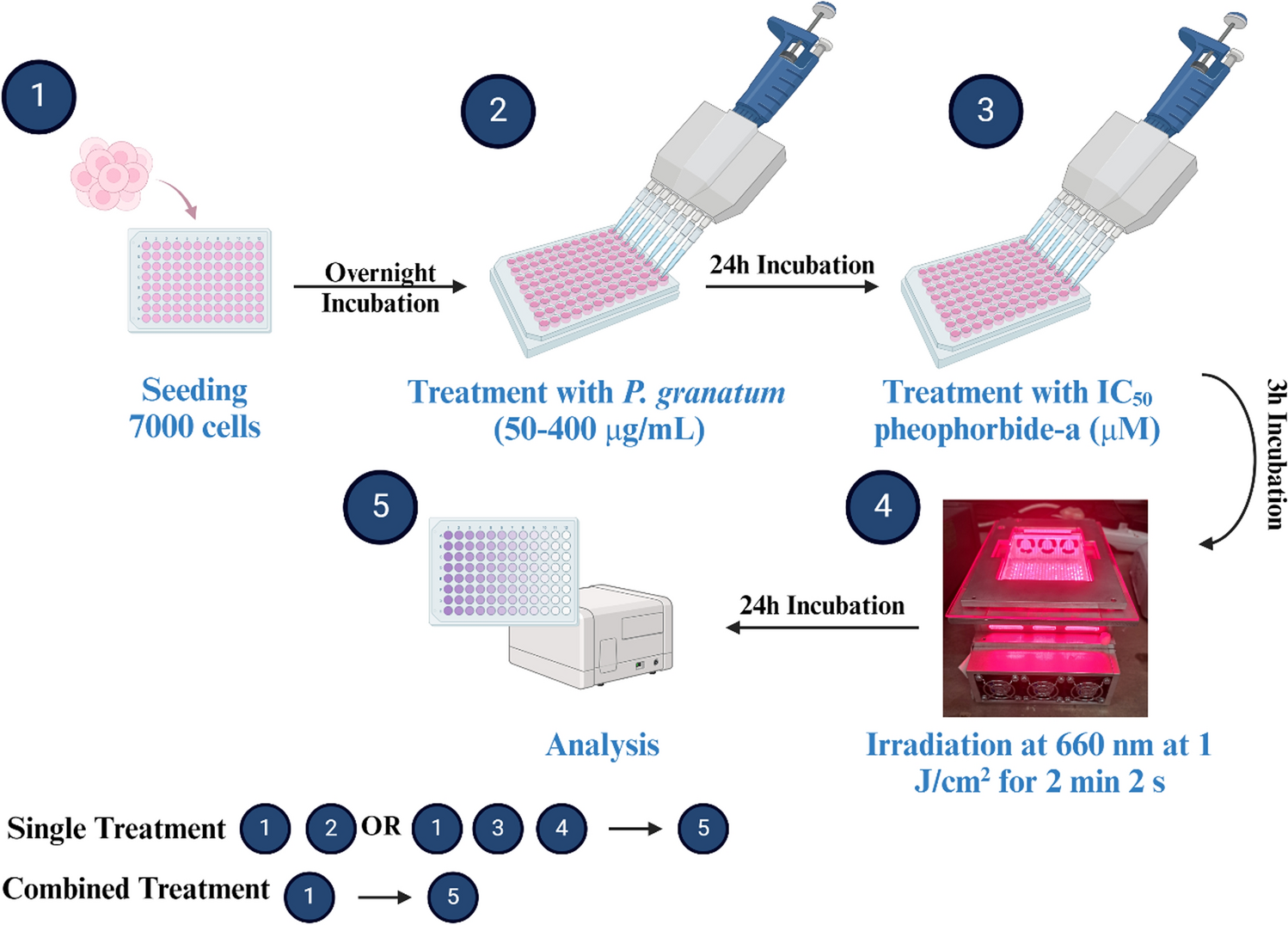
In our previous study, we investigated the phytochemical content and antitumor potential of South African Wonderful cultivar fruit (Punica granatum L.). The methanolic extract of arils with the highest phenolics and flavonoid content showed the most significant cytotoxic potential against MCF-7 induced by apoptotic cell death (Fakudze et al. 2024). Phytochemicals have demonstrated an ability to improve anticancer activity (Kim et al. 2014; Chen et al. 2018; Dou et al.2018) and overcome drug resistance in preclinical studies when combined with other therapies like chemotherapy. Other than the advantage of overcoming the induced negative effects, combination therapy with plant products enhances the therapeutic effect in a synergistic or additive way along with the reduction in delivery doses, drug dose toxicity, and lessening drug resistance (Aziz et al. 2023; Combination Anticancer Therapies Using Selected Phytochemicals—PubMed, https://pubmed.ncbi.nlm.nih.gov/36080219/). In this regard, the anticipated cross-resistance and overlapping negative effects of these compounds should be considered when creating a combination experimental model (Nikanjam et al. 2017). Interestingly, as per our knowledge this is the first study of its own for combination of P. granatum fruit extract with PDT.
Although in vitro studies cannot confirm in vivo biological activity, they do provide a rationale for using plant extracts for anticancer efficacy. However, caution must be taken when interpreting these findings, as several bioactive metabolites, which are frequently produced by intestinal or hepatic metabolism, may differ from those first found in the extract, and many chemicals utilized in vitro may not be accessible in vivo. Ellagic acid (EA), one of the major ingredients of pomegranate fruit extract, reached a Cmax of 31.9 ng/ml in human plasma 1 h after ingestion but was rapidly cleared from the bloodstream within 4 h, when administered with 180 ml of pomegranate juice containing 25 mg EA and 318 mg ellagitannins (ETs). This is due to EA's poor water solubility and tendency to accumulate in intestinal epithelial cells (Seeram et al. 2004). The same group in a different pharmacokinetics clinical trial reported a EA Cmax of 18.64 ng/mL, Tmax of 0.98 h, AUC of 50.07 ng/h/mL, and a half-life (t₁/₂) of 0.75 h in human subjects who consumed 180 mL of pomegranate juice containing 318 mg of ETs (Seeram et al. 2006). Another study reported the results following consumption of pomegranate extract by healthy human volunteers in the form of capsules containing 800 mg of extract per dose, providing 330.4 mg of punicalagins and 21.6 mg of EA. EA and its microbial metabolites urolithins A and B, their glucuronides, and dimethyl ellagic acid-glucuronide were detected in plasma, though punicalagins were not. These metabolites, likely formed in the colon by gut microbiota, appeared in plasma between 2 to 24 h post-ingestion, with inter-individual variability indicating variation in metabolism or prior dietary intake. Pharmacokinetics studies showed mean Cmax for EA in plasma was 33.8 ± 12.7 ng/mL, with a Tmax of 1 h post-ingestion (Mertens-Talcott et al. 2006). While the study conducted to study in vivo distribution of the pheophorbide A in tumor bearing rats, indicated its primary accumulation in the reticuloendothelial system, followed by the gut, lungs, and pancreas, with minimal presence in the skin. Notably, it showed prolonged tumor retention with a high tumor-to-pancreas ratio at 24 h, suggesting a favorable absorption spectrum and lower skin toxicity needed for PDT (Evrard et al. 1994). While in silico study with SwissADME predicted limited gastrointestinal absorption for the pheophorbide-a, likely due to its high molecular weight and substrate for P-glycoprotein, and demonstrated bioavailability of 56%, indicating that a substantial amount of the administered dose enters systemic circulation (Paul et al. 2024).
Since polyphenolic phytocompounds are known to absorb substantially in the 250–350 nm range, they are principally responsible for this UV-region absorption of the methanolic extract of the fruit (Anouar et al. 2012). The presence of this peak does not directly contribute to conventional PDT light activation. The extract's role in our combination treatment study was to work in concert with the PDT-activated agent, as a chemotoxic agent with a different mode of action than PDT to achieve a synergistic treatment outcome. Thus, the relevance of use of a potent PS like pheophorbide-a is due to its strong Soret band about ~ 410 nm and distinctive Q bands between 500 and 700 nm, with most notable absorption peak at 665–670 nm. This Q band is ideal for efficient ROS production and light penetration into biological tissues since it lies well within the therapeutic window (600–800 nm), which can improve the efficacy of treatment (Ethirajan et al. 2010).
Colocalization study results showed that pheophorbide-a, a well-known hydrophobic PS, accumulated in lysosomes and the ER but not in the mitochondria (Fig. 3) (RÖder et al. 2000). It is commonly known that receptor-mediated endocytosis causes more hydrophobic PS to bind with low-density lipoproteins and accumulate in endosomes and lysosomes (Rosenkranz et al. 2000; Benov 2014; Van Straten et al. 2017). Our results are consistent with studies conducted by Moret et al. whereby they showed that the pheophorbide-a was primarily located in the ER in MDA-MB-231 cells and induced apoptotic cell death following light irradiation (Moret et al. 2021). Notably, in contrast to other reports in the literature, we were unable to detect mitochondrial localization (Tang and Liu 2009a; Choi et al. 2014; Yoon et al. 2014). Treatment results are significantly impacted by the subcellular localization of PS. Upon PDT-induced oxidative stress, lysosomal membrane permeabilization results in release of cathepsins and other hydrolytic enzymes, and ER stress, all of which can cumulatively trigger apoptotic cell death. Studies have shown that, depending on the subcellular localization of the PS, different pathways are activated. For instance, lysosomal accumulation triggers p38 MAPK and mitochondrial apoptosis, while ER-localization disrupts ER homeostasis and activates Ca2⁺ signaling and the unfolded protein response via PERK. These primary signals can be amplified through caspase-dependent and independent pathways, with later-stage events potentially involving secondary pathways leading to mitochondrial cell death (Moserova and Kralova 2012). Delivery of therapeutic compounds to precisely the subcellular site of action leads to increased efficacy and reduced toxicity. Localization in the mitochondria and ER, which are crucial functional organelles in cells, can cause synergistic effects to cancer treatment through maximizing therapeutic indices, lowering the overall drug dose, and these multiple techniques alter the drug's initial mode of action, which then may lead to lowered chances of multidrug resistance.
Pheophorbide-a mediated PDT induced strong phototoxicity against MCF-7 cells at a light dose of 1 J/cm2, with IC50 and IC90 concentrations of 0.5 µM and 0.88 µM, respectively (Fig. 4). Further, the cell viability was assessed for combination therapy, whereby different methanol extract concentrations (0– 400 µg/ml) were combined with pheophorbide-a PDT at IC50 dose (0.5 µM, 1 J/cm2). As represented in Figs. 5 and 6, the effectiveness of pheophorbide-a mediated PDT showed a dose-dependent increase in combined treatment as compared to extract alone treatment. In the combination treatment, the IC50 dose was reduced to 129 µg/mL compared to single extract treatment of ~ 289 µg/mL and ~ 229 µg/mL at 24 and 48 h respectively (Fakudze et al. 2024). Interestingly an IC90 dose of 400 µg/mL was achieved with the combination dose, whereby 90% cell death was not observed with the methanol extract alone even at the highest concentration of 400 µg/mL within 48 h of the treatment period (Fakudze et al. 2024). A probable underlying mechanism for enhanced cytotoxicity in combination treatment can be explained as the synergistic interactions between the phytochemicals in the extract and the pheophorbide-a induced PDT. The phytochemicals present in the extract may sensitize cancer cells by modulating signaling pathways, whereby ROS generated during PDT directly harm cellular components, increasing cell death. When combined, these complementary processes have a stronger cytotoxic effect than when used as a single treatment. These results highlight the therapeutic potential of combination methods to improve efficacy at therapeutically feasible doses by overcoming the drawbacks of monotherapies, such as resistance development and partial cell death.
ROS generation is the central mechanism by which PDT exerts cytotoxic effects, leading to cancer cell death. Pheophorbide-a is a potent generator of singlet oxygen, making it highly effective as a PS in PDT (Wang et al. 2014). Thus, the photoactivation of pheophorbide-a leads to an increase in ROS, resulting in loss of cell viability. On the other hand, studies have shown that certain dietary phytochemicals demonstrate dual functionality, behaving as antioxidants or pro-oxidants depending on their concentrations and the oxidative state of the cellular environment (Fernando et al. 2019). Thus, based upon their activity, they can significantly influence the effectiveness of ROS-mediated cancer treatment at the molecular level. Thus, polyphenols, flavonoids, and anthocyanins present in Punica granatum aril extract could be acting as prooxidants under the concentrations used in this study (Fakudze et al. 2024). This redox modulation can amplify oxidative stress in cancer cells, thereby enhancing PDT efficacy.
The morphological analyses of MCF-7 cells using bright field microscopy provided additional evidence for the cytotoxic effect of single and combined treatments (Fig. 7 A-D). Both the single treatment and the combination treatment resulted in notable morphological alterations in MCF-7 cells relative to untreated control cells. The combination treatment group showed the highest cell toxicity as indicated by the majority of the free-floating cells, detached and rounded cells revealing the primary apoptotic features. The results were further confirmed with LIVE/DEAD fluorescent assay; compared to the single treatment, the combined treatment group (Fig. 7H) demonstrated a markedly higher percentage of dead cells, as indicated by fluorescent red nuclei, and nearly negligible green fluorescence, indicating a substantial decrease in cell viability.
To further, analyze the cell death mechanism in both single and combination treatment, several biochemical and molecular marker-based assays were carried out. FITC Annexin V/PI assay (Fig. 8) indicated induction of significant apoptotic cell death in all treatment groups. However, the combined treatment resulted in a significant increase in early and late apoptotic cell populations, of ~ 70% and ~ 18% respectively in comparison to all single treatment groups. These results are consistent with DNA damage detection assay with Hoechst 33,342 fluorescent staining as shown in Fig. 9. Whereby, as compared to untreated and single treatment groups, a greater number of cells in the combined treatment group displayed nuclear morphological characteristics such as DNA condensation and damage in dying cells, indicating apoptotic mode of cell death.
Several studies have suggested that different intracellular cell death pathways can be triggered in a cell type-specific manner by pheophorbide-a PDT. For example, pheophorbide-a mediated PDT-induced generation of ROS has been shown to induce apoptosis through the mitochondrial-mediated pathway in several human cancer cells such as uterine carcinosarcoma, pigmented melanoma, colon cancer, pancreatic cancer, hepatocellular, head and neck cancer cells and Jurkat leukemia (Hajri et al. 1999; Jin et al. 2000; Lee et al. 2004; Tang et al. 2006, 2009a, b; Chung et al. 2009; Tang and Zhang 2009b). While Yoon et al. showed that pheophorbide-a PDT leads to the activation of both autophagy and apoptotic pathways in the human breast, oral squamous carcinoma, non-melanoma, and melanoma skin cancer cell lines (Bui-Xuan et al. 2010; Ahn et al. 2013; Yoon et al. 2014). While in another study pheophorbide-a PDT was shown to cause G0/G1 cell cycle arrest in androgen-sensitive human prostate adenocarcinoma cell line (Gheewala et al. 2018). These results indicate that pheophorbide-a PDT may trigger different or overlapping pathways of cell death, depending on the cellular environment, its subcellular distribution, delivered PS concentration, and light dose as well as intracellular uptake limits.
Following the initiation of apoptosis by the different treatments in MCF-7 cells, alterations in the expression of the cytochrome-c, Bax (pro-apoptotic), Bcl-2 (anti-apoptotic), and caspase 8 and 9 proteins (Fig. 10) were found in order to understand probable molecular pathways that caused cells to undergo apoptosis. There are two main ways in which apoptosis is initiated: intrinsically though the mitochondria or extrinsically via death receptors, or DR. In contrast to the extrinsic pathway, which is triggered by death ligand binding to its appropriate receptor and subsequent caspase-8 activation, the intrinsic pathway is activated by cytochrome-c release from the mitochondria following inner mitochondrial membrane disruption and subsequent caspase-9 activation. Caspases-3 and -7, which are the ultimate agents of apoptotic cell death, are triggered by a sequence of signaling cascades that are initiated in these two pathways (Elmore 2007). It is commonly known that proteins belonging to the Bcl-2 family are essential for either initiating or blocking intrinsic apoptotic pathways (Qian et al. 2022). Combining treatments in this study resulted in enhanced levels of the proapoptotic protein Bax and a decline in the levels of the antiapoptotic Bcl-2 protein, which raised the ratio of Bax/Bcl-2. The increase in the Bax to Bcl-2 ratio causes the mitochondrial membrane to become porous, which allows cytochrome c release from the mitochondria and to enter the cytoplasm, where it triggers a series of caspase activation events that induce apoptosis (De et al. 2023). Notably, the increased Bax to Bcl-2 ratio and cytoplasmic cytochrome-c in the combined treatment was the highest with respect to single treatment groups. Further, the combined treatment additionally displayed a comparable chain of events denoted by increased cytochrome-c in the cytoplasm levels and increased caspase 8 and 9 activities.
The caspases, which are classified as either initiator or executioner caspases, include caspases -8 and -9 (initiators) and caspases-3 or -7 (executioners). During the leaking of mitochondrial cytochrome c into the cytoplasm, caspase-9 is triggered, while caspase-8 initiator triggers the extrinsic pathway (Elmore 2007). In our previous report, cells treated with methanol extract showed enhancement of caspase 9 levels, suggesting that the mitochondria-induced intrinsic pathway was involved in facilitating apoptosis. Furthermore, the amount of caspase 8 was also noticeably greater in treated cells than in untreated cells, indicating that the extrinsic apoptotic pathway was likely also involved (Fakudze et al. 2024). Interestingly, other than the heightened activity of caspase 9, pheophorbide-a mediated PDT at IC50 dose also showed a relative incline in caspase-8 activity (Fig. 9). This result is quite interesting compared to common characteristic of PDT-induced apoptosis which generally results in the quick release of cytochrome c from the mitochondria into the cytosol and the activation of caspase-9 pathway (Robertson et al. 2009). Pyropheophorbide-a methyl ester was reported to mediate PDT-induced apoptosis primarily via the mitochondrial caspase-9/-3 pathway intrinsic pathway (Matroule et al. 2001; Tian et al. 2006). While in another study, a PSMA inhibitor-conjugate of pyropheophorbide-a was shown to trigger apoptosis specifically through the caspase-8/-3 pathway (Liu et al. 2010).
The difference in the subcellular localization and PDT-generated ROS can be partially attributed to the difference in the activation of distinct apoptotic pathways (Liu et al. 2010). As pheophorbide-a was observed to localize partially in the lysosomes, the PDT-mediated disruption of lysosome and eventual release of cathepsins B and D, activation of proapoptotic protein Bid can be speculated to be involved in the activation of the intrinsic apoptotic pathway (Maharjan and Bhattarai 2022a). Furthermore, localization of pheophorbide-a in ER can also be responsible for PDT-induced ER stress to induce cell death, as also reported in other studies (Bui-Xuan et al. 2010; Gheewala et al. 2018).
Interestingly, the extent and type of ROS produced also determines the type of apoptotic caspase pathway involved. Certain studies have demonstrated that singlet oxygen mediated selective activation of p38 and caspase 8. This in turn signals the cleavage of Bid, which is further able to link to extrinsic and intrinsic apoptotic pathways. The active Bid protein causes depolarization of the mitochondrial transmembrane potential and releases cytochrome c, mediating the activation of downstream caspases (Zhuang et al. 2000; Salmerón et al. 2018). Furthermore, direct activation of the extrinsic apoptotic pathway can also be mediated by photogeneration of singlet oxygen which promotes the Fas receptor's ligand-independent oligomerization, which binds to the adapter protein FADD and causes the activation of caspase 8 (Morita et al. 1997; Zhuang et al. 2000; Maharjan and Bhattarai 2022b). Thus, pheophorbide-a with a high singlet oxygen quantum yield of ~ 0.69, is strongly speculated to be involved in activation of extrinsic apoptotic pathway along with the intrinsic pathway (Fernandez et al. 1997). Apoptosis in MCF-7 cells is mediated by caspases-9/8, 7, and 6 due to the lack of caspase-3 expression (Liang et al. 2001).
When compared to single treatment groups, the combined treatment group showed the highest activity of both caspase 8 and caspase 9 representing the involvement of intrinsic and extrinsic apoptosis pathways in MCF-7 cell death by this treatment (Fig. 10D). Our study showed strong synergistic/additive effects of using P. granatum extracts in combination with pheophorbide-a mediated PDT. There are very few reports on the combination of plant extracts or phytochemicals with PDT. One study demonstrated the lung cancer (A549) cell line's cytotoxicity, DNA damage, and apoptosis were all markedly and dose-dependently enhanced by the catechin combined with zinc phthalocyanine (ZnPcS4)-mediated PDT (Senapathy et al. 2020). Another study conducted with Dicoma anomala root extracts combined with ZnPcS4-mediated PDT to produce cytotoxic effects on lung (A549) and breast (MCF-7) cancer cells, which led to the upregulation of apoptotic proteins (p38, p53, Bax, caspase 3, caspase 8, and caspase 9) (Chota et al. 2022a; b). Aziz et al., also reported the synergetic effect of the combination of Ficus Carica and Ficus racemose extract with photosense-mediated PDT resulting in a pronounced cytotoxic effect, suppressed cell migration and cell apoptosis in rhabdomyosarcoma cells (Aziz et al. 2021; Aziz et al. 2023).
Although a direct comparison with conventional chemotherapeutic treatments such as doxorubicin or paclitaxel was not included in our current investigation, the literature indicates that these chemotherapeutic drugs, used as the first line of treatment for breast cancer, despite their high effectiveness, are frequently linked to dose-limiting toxicities and multidrug resistance(Crown et al. 2004; Okem et al. 2023; Kaveh Zenjanab et al. 2024). On the other hand, a combination of phytochemical-based treatment as used in this study provides a more effective treatment strategy that may result in selective cytotoxicity and less induced toxicities, at comparatively lower doses. Even though this is a preliminary study, the results demonstrate the therapeutic value of combining plant-based bioactive compounds with PDT as a complementary or alternative strategy, particularly when traditional treatments show limited responses or have serious adverse effects. The relative effectiveness, safety, and clinical transferability of this strategy will need to be confirmed by additional preclinical in vivo studies. Future studies are essential to confirm the therapeutic efficacy, pharmacokinetics, bioavailability, and safety profile of the combination therapy.


Comments (0)