Procedure.
A single male human investigator (the second author [ARN], aged 23 years) was provided with a 1-hour (approximately 8 am) and later a 2-hour float opportunity (approximately 7:30 pm) on 15 February and 26 March 2024 in a commercial float therapy center (City Cave, Glebe, Sydney, NSW, Australia). Brain and eye activity was recorded to show the feasibility and safety of recording EEG signals in a human floating in an Epsom salt solution. After the second nap opportunity, ARN also wrote a personal reflection on their experience before the EEG trace had been scored and has provided written consent to identification. As this was a technical feasibility study conducted with an author so that we could prepare a formal protocol, we did not have a formal human research ethical review for this initial test. We did receive formal governance approval to undertake the test from the Woolcock Institute for Medical Research in December 2023. The procedures were conducted according to the principles of the Declaration of Helsinki.
Setting.
Float REST therapy chambers are open pools approximately 40 cm in depth and about 2.0 m by 1.6 m. They are filled with saturated Epsom salt solution, which causes buoyancy in humans lying supine in the solution (specific gravity 1.23–1.28). The water is set to thermoneutrality (34.5 °C), and the room is sound and light isolated, although participants are able to control the lights via a waterproof button inside the pool. The room also has some floor area, which includes a shower. The Supplementary Materials include a photograph of the human floating in such a tank while the EEG is recorded as well as the connection to the protective housing around the EEG recorder.
EEG recording and processing.
Reading the existing literature on measuring EEG in floatation REST indicated that it might be possible to measure the frontal channels if we kept them from being submerged in the Epsom salt solution to prevent EEG bridging artifacts. As such, we built a prototype rig with a three-layer waterproof housing around the EEG recorder. The outer layer comprises a Styrofoam insulated box with a small hole drilled into it to allow entrance of the EEG leads. The middle layer inside this is a kitchen or fridge storage box suitable for soup, with a watertight seal through which the EEG leads are threaded. The inner layer is a thick plastic bag rendered watertight around the EEG leads using duct tape. The inner double layer withstood a 2-min full-immersion test. The technical specification brochure for the recorder also recommended that it be used below 35 °C,so we used it with an ice pack between the inner two layers to maintain a cooler temperature whilst avoiding moisture ingress to the inner layer. The innermost layer is also packed with paper towels to absorb any condensation. We used a setup montage of electrode placement on the face and forehead to keep the electrodes dry and free from interference from the Epsom salt solution (see Supplementary Figure). These precautions were a prerequisite of our research governance process to ensure the safety of our equipment and the safety of our participant.
We acquired the EEG using the Philips Alice PDx device and analyzed it in Alice G3 Sleepware 3.9.5 (Philips Respironics, Pittsburgh, PA, USA). We used standard silver chloride (AgCl) Grass EEG electrodes (Natus Medical Inc., Middleton, WI, USA). We used Ten20 as the conductive paste (Weaver and Company, Aurora, CO, USA) and Nuprep (Weaver and Company) for skin preparation. The EEG was recorded at derivations Fp1-Fpz and Fp2-Fpz [6,7,8]. A left and right electrooculogram (EOG) was recorded according to the American Academy of Sleep Medicine (AASM) placements, and these were referenced to Fpz [9]. Filter settings for recording were set at a high pass setting of 0.3 Hz and a low pass setting of 35 Hz, with a 50-Hz notch filter.
Unequivocal sleep was defined as the presence of a 30‑s epoch of stage 2 non-rapid eye movement sleep (NREM2) according to AASM criteria (v2.2). Confirmation of sleep spindles was via additional filtering within the EEG sigma frequency band between 11 and 16 Hz to visually isolate sleep spindle activity from the rest of the EEG.
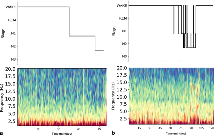
Quantitative EEG (qEEG) represents the composition of frequencies across the acquisition. Spectrograms, which are used to visually plot qEEG data, were generated using the YASA Python package using a multitaper method [10].
We defined the criteria for unihemispheric slow-wave sleep as the scoring of stage 3 NREM sleep (according to the AASM) in one hemisphere and wake in the other hemisphere in the same 30‑s epoch. We further hoped to confirm any hemispheric differences using quantitative techniques (such as the spectrogram), where obvious differences would be observed in lower frequencies between the left and right spectrograms.




Comments (0)