
Remember me
Searches on Pubmed on September 1 2025 and February 1 2026 were performed with the terms “segmental colitis associated with diverticulosis”, “segmental colitis” and “colitis AND diverticulosis.” Relevant titles were included in the initial literature review. Original research and literature focused on imaging were retained in the final references.
PathogenesisThe exact etiology of SCAD remains elusive, but it is widely accepted to be a multifactorial condition involving a complex interplay of mechanical and biological triggers [4, 10, 11]. One primary hypothesis is that fecal stasis within the diverticula facilitates microbial dysbiosis and prolonged contact between fecal material and the colonic mucosa, triggering a localized inflammatory response [11,12,13]. Additionally, mechanical mucosal prolapse resulting from intense colonic contractions and mucosal redundancy is considered a critical pathogenetic factor that leads to chronic mucosal trauma and subsequent inflammation [3, 9, 10].
Other researchers suggest that localized ischemia may occur in colonic segments experiencing high intraluminal pressure or changes in microcirculation, which further compromise the mucosal barrier [14]. This initial injury is often followed by a sustained immune-mediated response likely triggered by altered mucosal permeability, which increases exposure to intraluminal antigens and toxins [3, 14].
This persistent inflammatory state is further characterized by a genetically determined dysregulation of the host immune response, mirroring the pathophysiology of classical inflammatory bowel disease (IBD) through the overexpression of tumor necrosis factor-alpha (TNF-α) [3, 15]. Recent findings also implicate a reduction in beneficial bacterial species, such as those that produce short-chain fatty acids with anti-inflammatory effects, in sustaining the chronic inflammation observed in SCAD [13, 14]. Finally, some evidence suggests that SCAD might represent an idiosyncratic inflammatory manifestation of diverticular disease in genetically susceptible individuals rather than a separate disease process [12].
Clinical presentation and natural historyThe clinical triad of SCAD includes chronic diarrhea, crampy abdominal pain, and hematochezia, the latter of which occurs in up to 90% of patients [2, 5, 7]. While these primary symptoms are common, some patients may also experience constipation, tenesmus, or passage of mucus per rectum, though systemic signs such as fever, nausea, and weight loss are characteristically rare [11, 14].
In contrast to the more aggressive nature of IBD, the course of SCAD is generally favorable. Many cases resolve spontaneously without pharmaceutical intervention [1]. When medical management is required, patients often respond well to a limited course of antibiotics or oral 5-aminosalicylic acid (5-ASA) agents [16]. Severe, persistent, or refractory cases may necessitate more intensive therapy, including corticosteroids, biologic agents such as infliximab or adalimumab, or even segmental surgical resection for complications such as strictures or perforations [16]. While remission and relapse can occur, particularly in patients with ulcerative colitis-like endoscopic patterns, long-term follow-up studies have established no increased risk of developing colorectal cancer or adenomas in patients with SCAD compared to those with simple diverticulosis [1].
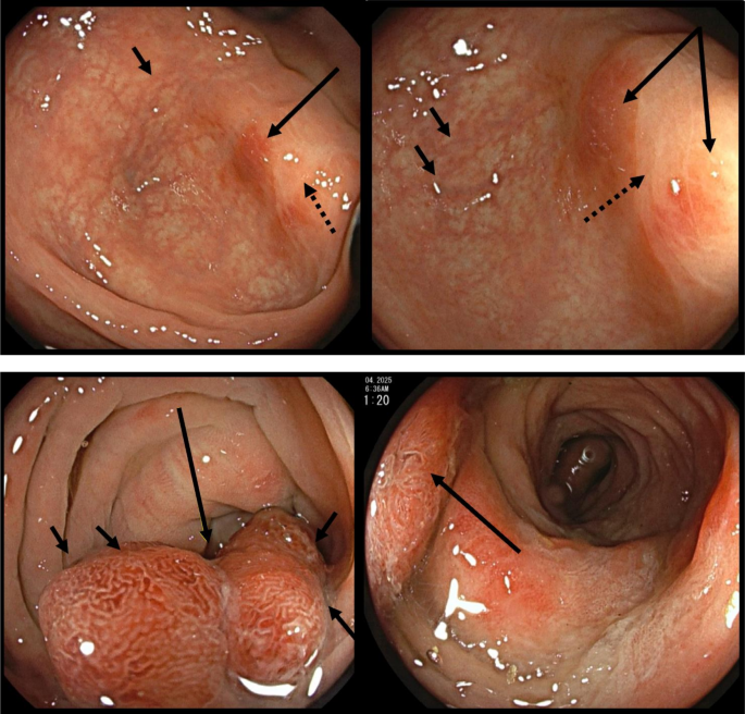
Diagnostic criteriaEndoscopy remains the gold standard for identifying SCAD [17] (Fig. 1). The unifying endoscopic features across its four recognized subtypes include inflammation restricted strictly to the diverticular segment, clear evidence of a normal-appearing rectum and proximal colon, and the characteristic sparing of the diverticular orifices themselves [4]. These endoscopic patterns range from mild crescentic fold lesions (Type A) and scattered aphthous ulcers to severe ulcerative colitis-like patterns (Type D), with the severity of the macroscopic pattern often correlating with the intensity of histological damage [10, 13].
Fig. 1
Select endoscopic features of SCAD. Top panels: Endoscopic images of mild SCAD. Focal regions of erythema (long arrow). Additional vascular congestion (short arrow) and loss of vascular pattern (dotted arrow). These findings are more common in mild types of SCAD. Bottom panels: Endoscopic images of moderate SCAD: Focal regions of erythema and vascular congestion (long arrow) with raised edges suggesting more profound mucosal edema (short arrows). Ulcerations and bleeding are not seen, as would characterize more severe types of SCAD
Comprehensive biopsy sampling is essential to exclude other clinical entities that can mimic SCAD, such as malignancy, classic inflammatory bowel disease (IBD), and drug-induced colitis [9].Common histological findings in affected segments include active inflammation markers, such as cryptitis and crypt abscesses, as well as chronic changes, such as goblet cell depletion and crypt architectural distortion [9] (Fig. 2). A summary of endoscopic and histopathologic findings is provided in Table 1.
Fig. 2
Select histopathologic features of SCAD. Top panel. Low-power image of diverticulum with intramural abscess (arrow). The presence of inflammation involving a diverticulum excludes SCAD. Second panel. Higher power image of intramural abscess, including numerous neutrophils, giant cells, and food particulate. No granulomas noted, which are a hallmark feature of Crohn’s colitis. Third panel. Background colon shows mild crypt distortion with irregular shape and size (labeled with stars), often seen in SCAD types B and D. Bottom panel. Background colon shows mild crypt distortion and Paneth cell metaplasia (labeled with star), often seen in SCAD types B and D
Table 1 Endoscopic and histopathologic features of SCADThe diagnosis of SCAD is best supported by a histologically normal rectal biopsy. This feature distinguishes SCAD from ulcerative colitis, which typically involves the rectum and lacks diverticula [16]. Furthermore, the absence of epithelioid granulomas typically distinguishes SCAD from Crohn’s disease, although distinguishing the two can be challenging when SCAD exhibits a chronic transmucosal inflammatory pattern [1]. Finally, clinicians must utilize laboratory analyses and detailed histories to exclude infectious etiologies, such as Salmonella, and iatrogenic causes, such as colitis induced by immunotherapy or NSAID use [18].
Imaging features of SCADConcordant imaging features are crucial for raising clinical suspicion of SCAD. Anatomically, SCAD involves an isolated segment of sigmoid or descending colon with associated diverticulosis. SCAD typically spares the right colon, transverse colon, and rectum. The most common CT and MRI findings include segmental wall thickening, sometimes with mural stratification, pericolonic fat stranding, and pericolonic free fluid. Penetrating disease, as manifested by perforation, fistulae, or sinus tracts, is less common but has been described, particularly with more severe subtypes [10]. Pneumatosis, frank pneumoperitoneum, and portal venous gas are rarelyseen in SCAD. As for most acute abdominal pathologies, CT remains the workhorse modality for diagnosing colonic pathologies and evaluating for SCAD. MRI can play a supporting role in illustrating colonic wall thickening separate from diverticula as well as pericolonic edema. MRI may also demonstrate diffusion restriction of the affected region of colon. Other findings include engorged vasa recta and the associated mesenteric vessels. Examples of common findings are shown in Figs. 3, 4.
Fig. 3
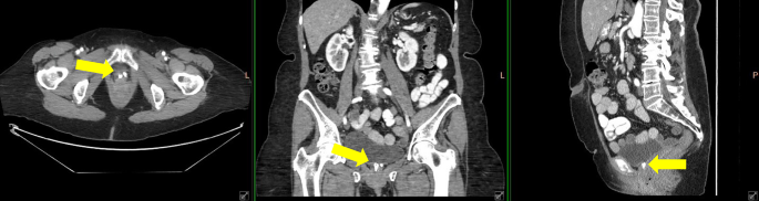
CT imaging features of SCAD. Top left panel. Thickened colon (long arrows) in a region affected by diverticulosis (short arrows). Arterial A and portal venous B phases are shown. The patient presented with hematochezia and was suspected to have ischemic colitis. Notably, no inflammation was noted about the diverticula. SCAD was eventually diagnosed after endoscopy and pathology. Top right panel. CT colonography images obtained for screening with persistently and segmentally thickened colon (long arrows) within a region affected by diverticulosis (short arrows). SCAD was eventually diagnosed after endoscopy and pathology. Bottom left panel. Sagittal contrast-enhanced portal venous phase image with colonic inflammation (long arrow) in an area affected by diverticulosis (short arrow) and characteristic rectal sparing (brackets). Bottom right panel. Maximum intensity projection contrast-enhanced CT pelvis in the coronal plane demonstrating dilated mesenteric vessels (brackets) adjacent to the inflamed sigmoid colon, the “comb sign.”
Fig. 4
MRI imaging features of SCAD. Top panel: Axial T1-weighted sequences demonstrating segmental thickening of colon (long arrow) in a region affected by diverticulosis (short arrows). Pericolonic fat stranding is noted (bracket). Second panel: Axial T2-weighted sequences demonstrating segmental thickening of colon (long arrow) in a region affected by diverticulosis (short arrows) without specific inflammation of the diverticula. The rightmost panel is applied with fat-saturation showing edema of the colonic wall (arrow). MRI can serve as an adjunct modality to CT in distinguishing inflammation of the colon from inflammation of a diverticulum. Bottom panel: Axial diffusion-weighted sequence with b-value of 800 (left) with paired apparent diffusion coefficient map (right) demonstrating diffusion reduction (bracket) at an inflamed segment of colon in a patient with SCAD. The concomitant ADC map is hypointense, indicating true diffusion reduction. Diffusion restriction of the affected wall is a key feature of SCAD on MRI, though not specific to that disease process
Generally, SCAD can be suggested when a patient presents with an isolated left-sided colitis in an area affected by diverticulosis. Inflammation should predominantly involve the colon wall and should not be centered on a single diverticulum, though secondary involvement of a diverticulum can occur [10]. Inflammation about a single diverticulum, particularly with abscess or perforation, more often suggests acute diverticulitis. Additionally, no rectal involvement should be seen. Other areas of colonic or even terminal ileal inflammation more likely reflect skip lesions in the setting of inflammatory bowel disease rather than SCAD. These findings are summarized in the flow chart seen in Fig. 5.
Fig. 5
Flow chart with algorithm for suggesting SCAD. Note that while exceptions exist, the majority of patients eventually diagnosed with SCAD demonstrate the following features on imaging. Lack of rectal involvement and colonic rather than diverticular inflammation are necessary to suggest the diagnosis. At best, SCAD may be suggested within a broader differential
Clinical overlap with other entitiesThe diagnosis of SCAD can be challenging due to the similar clinical and imaging features with other colonic pathologies, as well as its relative under-recognition in clinical practice. Common clinical features like abdominal pain and hematochezia overlap with those of diverticulitis and IBD. In a study with a final cohort of 75 patients diagnosed with SCAD, Urquhart et al. [10]. found that the most common clinical presenting symptoms were abdominal pain (33.3%), hematochezia (22.7%), diarrhea (14.7%), or two or more symptoms (24.0%). The mean age at diagnosis was 62.5 years (± 10.7 years), and 48% of patients were women. Of that cohort, approximately 61% had persistent or recurrent symptoms after first-line treatment, with 12 patients (16%) eventually diagnosed with inflammatory bowel disease. Of note, the average age of SCAD onset is similar to that of diverticulitis but significantly older than the typical age of onset for IBD, which is most often seen in the third or fourth decade [10]. Nausea, fever, weight loss, and leukocytosis are rarely observed [2].
Initial studies on SCAD considered it an atypical presentation of IBD rather than diverticular disease given its histopathologic findings, though with a relatively late onset and mild course, noting elevated levels of TNF-α, as seen in IBD [3, 15]. In a later prospective study of 27 patients with 5-year follow-up, a high rate of spontaneous resolution or resolution after a single course of treatment was observed. Unlike IBD, the rate of SCAD recurrence does not typically correlate with worsening endoscopic severity. However, patients who initially exhibit severe endoscopic patterns (types B and D) are more likely to experience a relapse or a more aggressive disease course. Importantly, SCAD shows no predilection for colonic adenomas or adenocarcinomas [17]. In fact, a more recent, larger prospective study examining the incidence of SCAD in a colonoscopy screening population suggested a slighter lower incidence of both adenomas and colorectal carcinoma in patients with SCAD [20]. Tursi et al. more recently highlighted in a limited case series that an endoscopic appearance suggesting SCAD is not enough to make the diagnosis – rather, negative histopathologic assessment of the right colon and rectum is needed in concordance with endoscopic appearance to avoid overdiagnosis of other entities, such as eosinophilic colitis or infectious colitis, which have typical pancolitic involvement of other segments of colon not seen in SCAD [18]. Collagenous colitis has been described in one case report as a mimicker for SCAD as well [21]. A rarer entity, diversion colitis, can be seen in segments of colon not involved in the fecal stream, such as those with ileostomies or colostomies, with overlapping endoscopic and histopathologic features [9].
With the rapid advancement of immunotherapies for solid tumors, the incidence of immunotherapy-linked colitis has increased in recent years. In a cohort of 16 patients treated with ipilimumab (Yervoy, Bristol-Myers Squibb, New York), 12 had diffuse colitis. In contrast, 4 had a pattern of colonic inflammation confined solely to diverticular regions, the so-called “SCAD pattern” of ipilimumab-associated colitis. Notably, the literature reports no endoscopic or histopathologic correlation [22]. In that study, patients with the SCAD pattern of ipilimumab-associated colitis presented with crampy abdominal pain, watery diarrhea, and hematochezia, rather than watery diarrhea alone, as seen in the diffuse pattern of colitis. Barina et al. subsequently describe a third colitis pattern involving the rectosigmoid region [23]. Given the suspected pathophysiology of classic SCAD, an altered immunologic milieu in the setting of ipilimumab-related SCAD may contribute to its development. While colitis associated with ipilimumab has been most frequently described, it would be reasonable to expect similar patterns with other immunotherapies, particularly those targeting PD-1/PD-L1.
Imaging overlap with other common entitiesAlthough patients often undergo imaging for abdominal pain, diarrhea, or hematochezia, nonspecific findings and overlap with other common entities make the diagnosis of SCAD particularly difficult. On CT and MRI, diverticulosis, IBD, and SCAD can present with segmental bowel wall thickening. The length of bowel segment involvement tends to be slightly longer in patients with SCAD (13.1 cm) compared to diverticulitis (9.8 cm) and Crohn’s disease (12.1 cm). While an inflamed diverticulum is much more commonly seen in acute diverticulitis, it does not exclude SCAD from the differential diagnosis. In one study, inflamed diverticula are present in approximately 39% of patients with SCAD, compared with 78% of those with acute diverticulitis. The same study shows moderate rates of pericolonic fat stranding and wall thickening (~ 63% and ~ 57%, respectively) and a low rate of peri-inflammatory free fluid (~ 12%). All three imaging features were seen at comparable rates compared to patients with both diagnosed acute diverticulitis and diagnosed Crohn’s disease. Moderate muscular hypertrophy occurs at comparable levels across all three disease processes. Interestingly, approximately 14% of SCAD patients demonstrate penetrating disease, such as a fistula or sinus tract [10] (Fig. 6).
Fig. 6
Mimickers of SCAD. Top panel. Muscular hypertrophy of the sigmoid colon. Segmental colonic wall thickening in an area affected by diverticulosis. SCAD was suspected, but subsequent biopsies failed to reveal active inflammation. Muscular hypertrophy is common in both SCAD and diverticulosis and cannot reliably be used to distinguish between the two entities. Second panel. Axial contrast-enhanced CT (left) in a patient after a hypotensive episode showing rectosigmoid junction wall thickening with a target-sign configuration of the bowel wall compatible with submucosal edema (arrow). Coronal contrast-enhanced CT (middle) in the same patient showing inflamed loops of rectosigmoid colon with mild surrounding fat stranding (arrows). Note the lack of colonic diverticula/diverticular inflammation. More posterior coronal contrast-enhanced CT image (right) in the same patient demonstrates an additional area of mild colonic inflammation at the level of the splenic flexure (arrow). Third panel. Axial contrast-enhanced CT images in a patient with a history of Crohn’s disease presenting with acute-onset abdominal pain (left and middle). There is an abscess in the rectouterine space (white arrow) with a developing fistulous connection between the sigmoid colon and the anterior rectal wall (dotted arrow). Numerous diverticula are present without significant peri-diverticular inflammation (black arrow). Coronal contrastenhanced CT image (right) in the same patient demonstrates muscular hypertrophy of the sigmoid colon (black arrow) and reactive pelvic lymph nodes (white arrow). Bowel wall thickening, penetrating disease (e.g., fistula or sinus tract), stricture, muscular hypertrophy, and peri-inflammatory free fluid are nonspecific imaging features shared between SCAD and inflammatory bowel disease. Note, ulcerative colitis will involve the rectum, while SCAD demonstrates rectal sparing. Crohn’s disease typically involves the terminal ileum and may demonstrate perianal sinus tracts or fistulae (but not in this case), while SCAD is generally isolated to a segment of sigmoid or descending colon. Penetrating disease is less common in SCAD than in Crohn’s colitis. Bottom panel. Axial contrast-enhanced CT images with enteric contrast showing an area of mass-like thickening (black bracket) and shouldering (short arrow) in the distal sigmoid colon corresponding to adenocarcinoma. Contained perforation with internal air and fluid contents is seen extending superiorly from the area of mass-like thickening (white bracket). While uncommon in the former, bowel perforation can be seen in SCAD and colorectal cancer
In rare instances, colonic strictures can develop in SCAD and, if involving a short segment, can occasionally mimic colonic adenocarcinoma [24]. In patients presenting with hematochezia, SCAD can mimic findings of ischemic colitis. Conversely, both colon adenocarcinoma and ischemic colitis can mimic SCAD in the setting of nonspecific clinical symptoms. Nearly all cases require optical colonoscopy to visually inspect the affected colon and histologically sample both involved and uninvolved segments. Perforation and imaging findings thereof, including pneumoperitoneum or abscess, are more specifically seen in complicated diverticulitis or IBD, but have been described in the setting of SCAD [25]. Table 2 compares the findings of SCAD with those of diverticulosis and IBD.
Table 2 Relative frequency of imaging features between common mimickers of SCADAlthough no studies specifically detail PET findings in SCAD, the affected segment would be expected to demonstrate hypermetabolism, closely mimicking the ‘SCAD pattern’ seen in ipilimumab-associated colitis, including vessel engorgement, wall thickening, and fluid-filled colonic distension [22, 23]. In such instances, clinicians must review the patient’s oncologic and medication history to distinguish immunotherapy-related colitis, as its treatment—cessation of the offending agent—differs from that of classical SCAD.
LimitationsEndoscopy and biopsy, not imaging, are the gold standard for the diagnosis of SCAD. At best, SCAD may be suggested in the impression of a report in the appropriate clinical setting, with further recommendations for direct visualization. Depending on the clinicians’ and pathologists’ familiarity with SCAD, a report may suggest taking endoscopic samples of uninvolved locations to exclude inflammatory bowel disease. Additionally, if a full workup has not been performed, recommendations for clinical correlation with drug history, stool studies, and laboratory testing may be appropriate.
Comments (0)