In this retrospective observational cohort study—the first to evaluate endoscopic performance in 82 CDH1 carriers within an Australian centre—we report real-world SRCC detection rates and surgical correlation over a 15-year period.
Most SRCC detections occurred during the first EGD, with substantially lower yield on subsequent procedures. This pattern suggests that most detectable lesions are identified early in the surveillance process [18]. The declining yield over time likely reflects both the underlying biology of CDH1-associated SRCC, characterised by intramucosal foci present from a young age, and the technical limitations of surveillance, whereby patients with repeatedly negative examinations may harbour microscopic lesions below current detection thresholds.
While diminishing yield after multiple negative examinations may suggest reduced incremental benefit of frequent surveillance [14, 20], the continued detection of SRCC in later procedures, including isolated cases at 5th and 6th endoscopies, supports the importance of maintaining surveillance programs for patients who opt against risk-reducing surgery.
With respect to the clinical context of our cohort: for much of the study period, guideline recommendations and counselling strongly favoured risk reducing gastrectomy, particularly following identification of SRCC [2, 22, 23]. Consequently, most EGDs were performed as preoperative assessments prior to risk reducing surgery rather than structured longitudinal surveillance. Over the past decade, however, data highlighting the ubiquity of pT1a lesions, the potential risk of overtreatment—especially in younger patients—and the low incidence of advanced cancer during surveillance have contributed to a more individualised, risk-adapted approach [16, 24, 25].
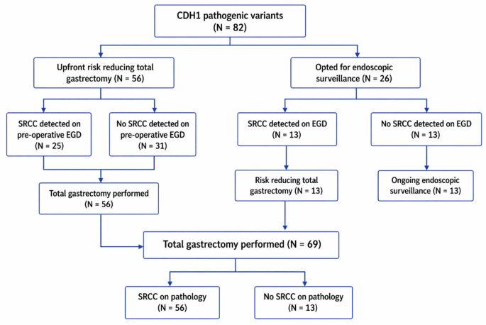
In the context of our cohort, among the 71 patients undergoing total gastrectomy, SRCC was present in 81.7% of specimens; however, invasive malignancy beyond pT1a was identified in only 2 patients (2.9%). Thus, although occult SRCC was common, progression to deeper invasive disease at the time of surgery was uncommon. Emerging surveillance cohorts have similarly shown low rates of progression to advanced gastric cancer, even among individuals with biopsy-proven intramucosal SRCC [16]. Our data support the growing recognition that many non-targeted, intramucosal SRCC lesions may be more indolent than previously assumed. These findings add to the contemporary perspective, with increasing acceptance of endoscopic surveillance as a reasonable option for selected patients rather than upfront risk reducing surgery.
With regards to endoscopic biopsy sensitivity, our cohort had an overall sensitivity of 67.9% for endoscopic SRCC detection, with a higher rate of detection in random biopsies than targeted biopsies (58.9% vs. 8.9%). The inherent biology of SRCC lesions make them difficult to detect as they typically manifest as microscopic foci without distinct macroscopic features, with previous models estimating that approximately 1700 biopsies would be required to achieve a 90% probability of detecting at least a single SRCC focus, highlighting the inherent limitations of endoscopic surveillance [23].
Studies utilising the earlier HDGC surveillance biopsy protocol have reported detection rates of 15–61% [9, 15, 18, 26, 27]. More recent approaches incorporating systematic and more extensive biopsy sampling have demonstrated improved diagnostic performance. The Lee et al. 16-year prospective study achieved 67.3% sensitivity in CDH1 carriers using a modified Cambridge Protocol [14] while Asif et al., demonstrated a 63% SRCC detection rate using the Bethesda protocol [16]. Our overall detection rate of 67.9% is largely consistent with the literature and reflects the contribution of systematic random biopsy protocols to the detection of occult SRCC.
However, the clinical utility of detecting isolated superficial pT1a SRCC lesions is uncertain. Current guidelines underscore that surveillance aims to exclude higher-risk disease rather than detect every microscopic focus (2). In this setting, random biopsies remain important in surveillance of patients deferring gastrectomy, although repeated sampling may also result in mucosal scarring that could potentially obscure subsequent SRCC lesions (18,28).
Furthermore, while intramucosal SRCC is frequently identified, the ability to distinguish lesions that will progress to clinically significant disease remains suboptimal, acknowledging that the risk of invasion is likely lower than previously thought and upfront risk reducing gastrectomy can safely be deferred or avoided. However, this is an important point in counselling of patients of the risk of developing an advanced diffuse gastric cancer. For some patients, the finding of an SRCC even if indolent is enough motivation to lead to risk reducing surgery and this is an important patient decision.
Interestingly, when calculated per biopsy, SRCC was detected in 1.2% of random and 7.8% of targeted samples. Although the per-biopsy yield of targeted biopsies was higher in targeted biopsies, these represented a minority percentage (2.4%) of total endoscopic biopsies performed; thus, the overall diagnostic contribution of targeted sampling remained limited. Of note, this finding is consistent with prior studies (18,29) which suggest that although targeted biopsies are fewer in number, they may contribute disproportionately to SRCC detection, while extensive random sampling yields a low per-biopsy diagnostic rate. Overall, these findings highlight the complementary roles of both targeted and random biopsy strategies, with careful mucosal inspection guiding targeted sampling, while systematic random biopsies may still detect otherwise occult lesions.
Macroscopic abnormalities have traditionally been considered potential sites of malignant change [2, 15, 16, 18, 28], particularly pale mucosal areas [14, 20]. In our study, while 4 of 24 (16.7%) documented pale areas detected SRCC, this did not reach statistical significance. Polyps demonstrated a significant negative association with SRCC detection, consistent with prior studies reporting SRCC detection rates within polyps of 0–1.8% [14, 16, 20], without a clear significant positive association. This is consistent with our findings and corroborates that SRCC are rarely found in polyps.
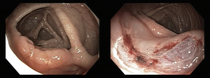
Importantly, the two cases of invasive disease (> pT1a) in our cohort were associated with visible mucosal abnormalities and were recognised as suspicious at the time of endoscopy, with representative endoscopic images shown (Fig. 3). This emphasises that, while detection of occult intramucosal SRCC may be imperfect and of uncertain clinical consequence, careful endoscopic inspection remains critical for the identification of lesions with features suggestive of more advanced disease.
The low prevalence of H. pylori observed in our cohort is broadly consistent with the limited published HDGC surveillance literature (14,16,19), which has suggested that CDH1 carriers may be at lower risk of H. pylori infection. This difference should also be interpreted in the context of ascertainment bias and age, as non-CDH1 comparator cohorts undergoing endoscopy are typically older and symptomatic, whereas CDH1 carriers are generally young and undergo screening or surveillance endoscopy.
Postoperative morbidity post gastrectomy was considerable, with 30.4% of patients requiring further intervention within 90 days—most often endoscopic dilatation for anastomotic stricture, a commonly demonstrated complication in contemporary HDGC series post total gastrectomy [29, 30].
Two patients (2.9%) developed an anastomotic leak and there were no procedure-related deaths, consistent with previous series reporting uncommon major surgical complications and procedure-related death [9, 31,32,33,34]. Patients should be counselled accordingly that although mortality is rare and invasive malignancy beyond pT1a is infrequent, up front gastrectomy remains a life-altering operation associated with meaningful functional consequences.
With respect to anatomical distribution, some studies have described a distal predominance of SRCC particularly within the antrum and transitional zone [14, 20, 23]. In contrast, our cohort showed a higher frequency of proximal involvement, with 39.4% of lesions in the fundus and 33% in the antrum, with some literature having a similar finding of fundus-predominant distribution in their cohorts [37], suggesting that proximal involvement may be more common than previously recognised. This variation may reflect true biological heterogeneity or differences in pathological sectioning and reporting. Our findings support the importance of systematic sampling across all gastric regions during endoscopic surveillance and in-depth examination of the entire surgical pathology specimen.
Gastric cancer risk assessment in CDH1 carriers identified through lobular breast cancer remains an area of evolving clinical importance. Increasingly, CDH1 carriers are identified through breast cancer–focused genetic testing [35], raising important questions about gastric cancer risk in such families [7, 36]. In our cohort, 39% of patients reported a family history of LBC and 10.7% had a personal history of LBC.
While SRCC was frequently identified in patients with a personal history of LBC or LCIS, no cases of invasive disease beyond pT1a were observed, suggesting that breast phenotype alone did not predict more advanced gastric pathology. These findings suggest that while gastric cancer risk remains clinically relevant in breast-presenting CDH1 families [38, 39]. LBC phenotype may not correlate with an increased risk of invasive gastric cancer. Accordingly, LBC patients and families should undergo gastric risk assessment and structured surveillance counselling, with decisions regarding prophylactic gastrectomy individualised according to family history, age, and patient preference [2].
This study has several important strengths. With 82 CDH1 carriers, 136 EGDs and 69 gastrectomies performed over 15 years, this represents a substantial single-center surveillance cohort providing sufficient data for meaningful analysis. The high proportion of patients proceeding to gastrectomy allows robust pathological correlation, and the 15-year span reflects evolving clinical practice from routine early risk reducing surgery toward more individualised surveillance strategies.
Several limitations should be acknowledged. As a retrospective single-centre study, our findings may not be generalisable. Many patients underwent only one EGD prior to up front risk reducing surgery, limiting comparability with contemporary structured surveillance cohorts. Use of surgical pathology as the reference standard may overestimate the clinical significance of microscopic SRCC, which may not inevitably progress to invasive cancer. Additionally, histopathological analysis of gastrectomy specimens may have been insufficient at times. Finally, relatively short surveillance follow-up (median 14.5 months) restricts assessment of long-term progression risk in patients managed non-operatively.


Comments (0)