
Remember me

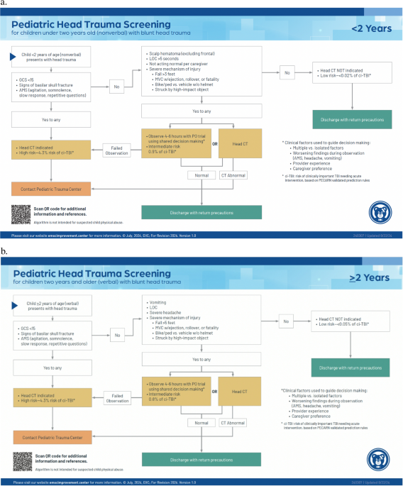

Head trauma is a leading reason children seek emergency care. Injuries can range from soft tissue contusions, cephalohematomas, and concussions to intracranial hemorrhage and severe traumatic brain injury. Evidence-based clinical decision tools have been developed to identify those requiring intervention with clinically significant head injuries, for which early recognition is paramount. Among the most commonly used is the PECARN prediction rule for head trauma [14, 18, 26]. PECARN published a large, multicenter prospective cohort study of over 42,000 children with minor blunt head trauma (Glasgow Coma Scale [GCS] score ≥ 14) to derive clinical decision rules for identifying children at very low risk of clinically important traumatic brain injury (ciTBI). These rules were designed to reduce the use of cranial CT imaging [4]. The PECARN rule uses age-stratified criteria (under 2 years or 2 years and older) that include clinical signs and symptoms, as well as the mechanism of injury. Children who meet the low-risk criteria per PECARN have a < 0.05% risk of ciTBI. The rule has 100% sensitivity and 100% negative predictive value, allowing for safe observation and avoidance of CT imaging [26]. The PECARN head trauma decision guide has been validated in various clinical settings and is supported by several societies in their Choosing Wisely recommendations [3, 14, 18,19,20]. Fig. 1a provides imaging recommendations aligning with PECARN for patient under 2 years old and 1b for those 2 years and older.
Fig. 1
EIIC Pediatric Head Trauma Screening Algorithm. The references PDF resources are free and open-access material and are licensed under the Creative Commons Attribution-NoCommerical-NoDerivates 4.0 International (CC BY-NC-ND 4.0). Image source:https://emscimprovement.center/education-and-resources/peak/multisystem-trauma/imaging/
Cervical SpineCervical spine injury occurs in 1–2% of pediatric trauma patients [27]. It can lead to significant morbidity and mortality, and its detection needs careful consideration. While there are well-validated clinical decision tools developed to assist with the evaluation of the adult cervical spine, few include a robust number of pediatric patients, particularly the very young [11, 28]. When the Canadian C-spine Rule (CCR) and National Emergency X-Radiography Utilization Study (NEXUS) were applied to children, they did not perform as well as in adults [29]. Many adult imaging guidelines do not account for the developmental and physiologic differences in the pediatric spines of younger children who have an increased risk of ligamentous and atlanto-axial injuries [28, 30]. The application of pediatric-specific protocols is warranted to evaluate the cervical spine in children, particularly with increased risk of ligamentous injury without fracture, compared to adults [31]. Additionally, given the radiosensitivity of the thyroid, a single cervical spine CT in childhood can increase the lifetime risk of thyroid cancer by 78% [8, 32].
After the Canadian C-Spine Rule and Nexus Criteria were implemented in the adult population, preliminary work showed these adult criteria were limited in their application to the pediatric population [29, 33]. In 2013, Rozzelle led a group to develop evidence-based recommendations for evaluation of the pediatric cervical spine, providing criteria to defer imaging in the asymptomatic, well-appearing child, called the PEDSPINE trials. Their recommendations evaluate children based on age group (under 3 years or 3 years and older), mechanism of injury, physical exam, and clinical symptoms, providing recommendations of radiography, high-resolution CT imaging, or magnetic resonance imaging (MRI) based on set criteria [30, 34].
Understanding developmental and physiologic differences in pediatric spines between ages, children < 3 years with a high-risk mechanism of injury are excluded from the defined exclusion criteria. Later, the Pediatric Cervical Spine Clearance Working Group convened experts and developed an algorithm for evaluation of the pediatric cervical spine, which was endorsed by several societies. Their recommendations are based on evaluability for a given Glasgow Coma Score (GCS) as well as anticipation for improvement of GCS. For lower-risk patients with GCS > 8 or those likely to improve, x-ray is the initial screening modality, with CT being recommended for those with a higher risk of c-spine injury (GCS ≤ 8). Special considerations of this algorithm included children suspected of abusive head trauma and those with high-risk mechanisms of injuries, such as high-risk motor vehicle collision, diving, clothes-lining, and axial load [27, 35].
Following much of this recent literature, PECARN updated its prediction rule to stratify patients into three risk categories [15, 36, 37]. Those with no risk factors, like previously mentioned work, do not need imaging of their cervical spine for clearance with a 0.2% risk of injury. Patients with classification and regression tree (CART) analysis-derived risk factors have a 2.8% risk of injury, and a plain x-ray should be considered. In patients who are high-risk, such as those with a GCS ≤ 8, abnormal airway, breathing, or circulation, or focal neurological deficits on exam, CT should be considered. Determination of the recommendation of CT imaging was based upon the risk of radiation exposure-induced malignancy and the benefit of identifying an injury. With this stratification, when applied to the original study population, more than 50% of the children undergoing CT could have avoided CT imaging without missing any clinically relevant injuries. In the adult population, the Eastern Association for the Surgery of Trauma (EAST) guidelines for cervical spine clearance recommend removal of the cervical collar after a negative CT alone [38]. This practice does not have sufficient evidence to be applied to the pediatric population. While the rate of operative injuries missed by CT use alone is very low, many unstable ligamentous injuries are treated with a rigid collar and not surgery in pediatric patients and are not accounted for in studies [34, 39].
While CT is commonly used in the evaluation of the adult trauma patient, this exposes a child to significant radiation. Application of clinical evidence-based guidelines, such as Fig. 2, can assist in identifying children at low risk, and imaging can be deferred, assisting with imaging decisions on increased risk patients. Recognition of children exceeding your center’s capability is paramount, and imaging of the cervical spine can be deferred until evaluation at a pediatric trauma center or in conjunction with consultation with the accepting center.
Fig. 2
EIIC Pediatric Cervical Spine Injury Screening Algorithm. The references PDF resources are free and open-access material and are licensed under the Creative Commons Attribution-NoCommerical-NoDerivates 4.0 International (CC BY-NC-ND 4.0). Image source: https://emscimprovement.center/education-and-resources/peak/multisystem-trauma/imaging/
Given injury location and mechanism of injury, children, like adults, are at risk for blunt cerebrovascular injury (BCVI). A variety of BCVI screening tools, such as the Memphis, Denver, Utah or McGovern, have been applied to pediatric patients. However, these tools are not as effective in children as they lack both sensitivity and specificity in children, subsequently causing injuries to be missed and too many unnecessary scans to be performed [40,41,42,43,44]. Prudent consideration of CT angiography of the neck in a child with risk factors for BCVI can decrease additional radiation exposure in children [45].
ThoraxExpedited diagnosis of life-threatening thoracic trauma is incorporated in the primary survey of the injured child with a significant mechanism of injury. The screening test most often utilized is the chest x-ray (CXR). This can aid in the diagnosis of pneumothorax, hemothorax, chest wall trauma, and pulmonary contusions. The mediastinum can be evaluated for widening, which can be a sign of cardiac or great vessel injury. Such a finding can be seen in high-risk patients who present after a decelerating mechanism with a blunt aortic injury. While a reliable diagnostic adjunct, discerning between a mediastinal widening and a normal thymus in young children may be difficult and misleading. There are certain situations where CXR should be supported by additional imaging. For example, when the CXR indicates an enlarged cardiac silhouette, a pericardial ultrasound is an important adjunct to accurately assess for hemopericardium. The CXR involves a relatively low radiation exposure when compared to CT of the thorax [46, 47]. Ultrasound has been examined as a potential means of reducing radiation exposure while providing rapid diagnosis. In the pediatric population, ultrasound has not shown the same reliability for pneumothorax as seen in the adult population, making it challenging to adopt broadly [48]. The algorithm provided by the Emergency Medical Services for Children Innovation and Improvement Center (EMSC EIIC) displays these decision points in choosing imaging modalities in patients with thoracic trauma in Fig. 3.
Fig. 3
EIIC Thoracic Trauma Screening Algorithm. The references PDF resources are free and open-access material and are licensed under the Creative Commons Attribution-NoCommerical-NoDerivates 4.0 International (CC BY-NC-ND 4.0). Image source: https://emscimprovement.center/education-and-resources/peak/multisystem-trauma/imaging/
Chest CT for routine screening of pediatric trauma patients for thoracic injuries should not be routinely used. Additionally, in the absence of a high-risk mechanism, such as a deceleration injury, a CTA is not indicated [11, 49]. The risk of radiation exposure compared to the benefit of diagnosing a condition not identified on CXR that would require an intervention has been called into question. Chest CT has been shown to diagnose more injuries than CXR alone, but none of these additional diagnosed injuries required specific treatment [47, 50,51,52]. The decision to order a CT scan for a pediatric patient should be considered in patients with both an abnormal mediastinum on CXR and a vehicle-related mechanism of injury [50, 53, 54]. If the patient is being evaluated at a center that will likely transfer children with chest injuries, consultation with the referral center prior to obtaining advanced imaging is prudent. The constant question of whether to CT scan a child is due to concerns of the long-term effects of additional radiation exposure, as previously described [8, 22, 55].
Abdomen/pelvisAbdominal trauma can be present in up to 25% to 33% of pediatric trauma patients and is the most common cause of a missed fatal injury. Most injuries are from blunt abdominal trauma, with 95% able to be managed nonoperatively. For those with rare penetrating injuries, operative intervention is more frequently required [56, 57].
There are two general approaches to clinical prediction rules for blunt intra-abdominal injury that are primarily differentiated by the need for laboratory values. Clinical prediction rules for blunt abdominal trauma have been in use since the early 2000s and have evolved over the subsequent two decades [58]. We will discuss the two most commonly used algorithms and propose a unified stepped-up approach that incorporates the benefits of both algorithms while minimizing overuse of CT and missed injury.
The PECARN abdominal injury rules have criteria based solely on findings from the patient’s exam and history. It should be noted that the prospective PECARN intra-abdominal injury study was designed to evaluate a prediction rule incorporating laboratory values, but the frequency of lab draws in the observational study was sufficiently low as to limit the utility of the laboratory values in the analysis [59]. The PECARN abdominal injury rules include evidence of abdominal wall trauma, GCS, abdominal tenderness, evidence of thoracic wall trauma, abdominal pain, presence of breath sounds and vomiting [59]. The criteria were designed to highlight which pediatric patients would likely be low risk for having sustained an abdominal injury that would require an intervention, rather than as a tool to direct whether CT imaging should or should not be obtained. As some of the included variables are subjective, they may result in the overuse of CT scans. The use of these criteria results in a sensitivity of 100% and specificity of 47.2% for clinically important intra-abdominal injuries if the criteria are used to mandate CT scanning [12].
The Pediatric Surgery Research Consortium (PedSRC) rules were derived from a subgroup analysis of the PECARN observational dataset that specifically included the children who had labs drawn, and are based on clinical, lab, and imaging findings. The PedSRC rules included: AST > 200 U/L, abnormal abdominal exam, abnormal CXR, abdominal pain, and abnormal pancreatic enzymes. These criteria result in a sensitivity of 98.4% and specificity of 38.1% for intrabdominal injury and a sensitivity of 100% and specificity of 34.7% for intra-abdominal injury requiring intervention [60].
Laboratory tests can help guide care, especially for those suspected of having an intra-abdominal injury. Various cutoffs of AST/ALT to prompt further imaging have been proposed. When there are physical exam findings concerning for intra-abdominal injury, the values of an AST > 200 IU/L or ALT > 125 IU/L should be followed by a CT scan of the abdomen to limit missing clinically significant injuries [59]. While low-grade hepatic injuries are not clinically significant, they offer value in the forensic adjudication of child physical abuse, and the much lower cutoff of 80 IU/L is utilized for such a purpose [61]. Lipase is often checked as a screening test in presenting trauma patients, and its elevation is highly sensitive but not predictive of the grade of a pancreatic injury [62]. When lipase is abnormal, further investigation with an abdominal CT is recommended [60]. Urinalysis is often performed for suspicion of urologic injury in trauma. Historically, identification of microhematuria on urinalysis resulted in further imaging. Multiple studies have now shown that microhematuria does not lead to improved identification of intra-abdominal injuries [63, 64]. Hematuria in the hemodynamically stable patient is further investigated with a split-bolus contrast CT abdomen and pelvis, which captures arterial, venous, as well as renal opacification from a single radiation dose [65].
The EMSC EIIC algorithm (Fig. 4) uses a two-phase approach [66]. High-risk factors get a CT scan and likely transfer to a pediatric trauma center. In the absence of high-risk factors, we recommend screening with the PECARN abdominal rules. If they screen negative, then recommend a six-hour observation period and discharge home with return precautions. If they screen positive by the PECARN rules, then labs and re-assessment are recommended. If the labs and CXR are normal and there is no abdominal tenderness, the patient can be observed for six hours and discharged home after a PO trial, and return precautions are given.
Fig. 4
EIIC Abdominal Trauma Screening Algorithm. The references PDF resources are free and open-access material and are licensed under the Creative Commons Attribution-NoCommerical-NoDerivates 4.0 International (CC BY-NC-ND 4.0). Image source: https://emscimprovement.center/education-and-resources/peak/multisystem-trauma/imaging/
With increased concerns regarding radiation exposure and improvement of ultrasonography, the Focused Assessment with Sonography for Trauma (FAST) is often utilized in the setting of pediatric trauma patients [56, 57]. It was developed as a hemorrhage localizing study in unstable patients and should continue to be utilized within this context. Its use as a screening tool in hemodynamically stable children is controversial, with data from a randomized control trial suggesting there is no benefit in identifying injuries and may even increase the use of unnecessary CT [67]. Thus, we do not recommend its use in the hemodynamically stable patient in the consideration of who needs a CT. Contrast-enhanced ultrasound is a promising modality, but it is a time-consuming study, requires specialized expertise, and is operator-dependent.
Overall, for pediatric blunt abdominal injury, well-validated clinical criteria provide an effective screen for further injury. These recommendations must be considered in the context of a center’s capabilities. In centers that do not have pediatric surgery support, the abdominal CT should be used to screen children (if indicated) to facilitate potentially discharging home from the ED. Abdominal CT based on the mechanism alone without other clinical risk factors is not necessary.
Whole Body ImagingWhole body CT (WBCT) imaging is a common practice in the adult population, particularly in those suspected of having multi-system injuries or altered mental status after injury. While it has been shown to decrease the rate of missed injuries, improve mortality rates and time to diagnosis in the adult population, the same level of benefit is not provided in the pediatric population due to the known risks of radiation, and consequently, body-region specific imaging is primarily used in this population [68, 69]. While certain indications do exist that would prompt a whole body CT scan of pediatric patients, these patients are more likely to undergo this type of workup in adult vs. pediatric trauma centers [11].
While for the aforementioned reasons, WBCT has not been widely applied in pediatric trauma patients, there are situations where they do provide significant benefit. A consensus on the exact indications for WBCT has not been established even in the adult population, but it is likely to provide the most benefit in patients with a limited physical exam, with whom the timely diagnosis of an injury would lead to immediate intervention or change in management [70, 71]. This may include patients with high-risk mechanisms and impaired neurologic status with decreased GCS or concern for cervical spinal cord injury on exam.
Comments (0)