
Remember me
Two-photon microscopy (2PM) has emerged as a key enabling technology for fluorescence imaging in complex biological specimens [1–3]. The quadratic dependence of excitation on intensity and the use of near-infrared light favour signal generation only close to the focal region [2–5]. Thereby, reducing much of the out-of-focus background that limits conventional widefield imaging in weakly scattering tissues [2, 6–8]. Since its adoption in the life sciences, the 2PM dominant implementation has been driven by laser point-scanning 2PM, which has enabled diverse deep-tissue studies at cellular resolution [5, 9]. Extensive work [10, 11], has explored both the biological applications and the technical behaviour of point-scanning 2PM, highlighting trade-offs between speed, sensitivity and photodamage. In response to these limitations, a variety of parallel and multi-beam strategies [10–13] have been proposed [12]. Those multiplexed concepts have shown that two-photon emission can be recovered without scanning a single diffraction-limited spot across the sample, opening a route to higher frame rates and simplified detection geometries. As well, in close analogy with the evolution from confocal scanning to camera-based widefield [14] detection in linear microscopy, two-photon excitation has also been implemented in widefield and scan-less formats [2]. In this implementation, temporal focusing (TF) has become the most widespread solution for this task.
Although femtosecond excitation is commonly employed in 2PM, pulse duration alone does not determine the feasibility or efficiency of nonlinear signal generation. For a pulsed source, the detected two-photon fluorescence depends jointly on average power, pulse energy, repetition rate, pulse duration, focusing conditions, excitation wavelength, and fluorophore properties [15–18]. Consequently, multiphoton imaging has also been demonstrated with picosecond and nanosecond sources when sufficient pulse energy is delivered, commonly through operation at reduced repetition rates. Picosecond excitation can provide two-photon fluorescence and second-harmonic generation comparable to femtosecond excitation while being less sensitive to chromatic dispersion [16]. Nanosecond and sub-nanosecond supercontinuum sources have likewise enabled multicolour two-photon fluorescence, harmonic generation, and multimodal imaging in biological specimens [17, 18]. These results show that the operating regime is defined by the combined temporal, spectral, and energetic characteristics of the source, rather than by pulse duration alone.
A different constraint arises when excitation is distributed over an extended sample region rather than concentrated into a diffraction-limited scanning focus. Under scanless or patterned illumination, the available pulse energy is shared across a larger area, reducing the local instantaneous intensity available for nonlinear excitation. TF has therefore been widely employed to recover efficient and axially localized excitation by spectrally dispersing the pulse and recompressing it only near the sample plane [19–22].
Beyond the optical requirements of extended-area excitation, replacing point-by-point scanning with projected illumination patterns also changes how spatial information is acquired. In single pixel imaging (SPI) spatial information is encoded through a sequence of known illumination patterns, while the total fluorescence generated by each pattern is integrated by a non-spatially resolving detector. The image is subsequently reconstructed by correlating the resulting scalar measurements with the projected patterns. This architecture is particularly relevant for multiphoton microscopy because it combines extended-area patterned excitation with sensitive bucket detection and enables Hadamard-based and compressive acquisition strategies.
In that way, only the dispersive element conjugated plane nonlinear excitation is recovered over a widefield-excitation of view. However, while this concept is powerful and has been combined with structured illumination [20], spatial light modulators [8] and digital micromirror devices (DMDs) [23], it entails substantial practical overhead. Strong spatiotemporal dispersion must be introduced and precisely compensated, the diffracted angular wavelengths must be relayed accurately through the system, and the full cone of temporally dispersed rays must pass through the objective pupil [21, 22, 24, 25]. In practice, these requirements are fulfilled with high numerical aperture (NA), and large back-aperture objectives [26, 27]. The cost of those specialized elements rounds the 20.000 USD, being in the level of the budget for a scientific grade microscope. Additional to the budget constraint which optics handling additional complexity due to their short working distances related to their high NA. As a result, widefield 2PM systems are often pushed into a price bracket dominated by premium high-NA optics, specialized ultrafast sources and complex motion controlling systems, which stills leaves clear room for experimental simplification and broader accessibility [27].
Even when widefield nonlinear excitation is successfully implemented, imaging depth and robustness remain limited by the optical properties of the specimen. The main advantage of 2PM lies in the ability to localize excitation deeper in weakly scattering media, but the emitted fluorescence must still propagate back through the turbid layer [1]. Multiple scattering, aberrations and angle-dependent transmission distort the wavefront and redistribute photons away from ballistic paths, so that camera-based widefield detection again collects a mixture of useful signal and scattered light. To mitigate this constraint, patterned-excitation schemes that do not rely on spatially resolved detection have been introduced [12, 13, 28, 29]. The TRAFIX [12] implementation, for instance, employs SPI together with orthonormal pattern projection and computational reconstruction. Showing that a relatively small set of structured two-photon measurements can substitute for dense point scanning while SPI is less vulnerable to degradation of the emitted wavefront [12, 29–31]. This shows that SPI can deal with the widefield collection problem, nevertheless the reported integrations must still be constrained to complex experimental need of TF implementation [13].
In this work, we present an experimental approach to widefield-excitation two-photon single-pixel microscopy (2P-SPM) significantly simplifying the optical setup avoiding the complexity of TF while retaining the core advantages of SPI strategies, including compressive sensing (CS) and robustness in weakly scattering media [30–35]. Binary Walsh-ordered and scrambled Hadamard patterns are projected onto the specimen, the total fluorescence associated with each pattern is measured with an avalanche photodiode (APD) bucket detector, and images are recovered by correlation-based or compressive reconstruction [13, 31, 32, 36]. However, reported multiphoton SPM implementations remain tied to TF modules [12, 13, 27, 28] and sub-200-fs laser sources, because the dispersive patterning elements broaden the pulses and reduce nonlinear excitation efficiency unless elaborate temporal recompression is engineered. This coupling between temporal shaping, pattern projection and ultrashort-pulse management has so far limited the experimental simplicity and accessibility of widefield 2PM in the single-pixel regime [27]. In the present work, these constraints are relaxed by demonstrating widefield 2P-SPM without TF, driven by an industrial laser at 1064 nm delivering 300-fs pulses and implemented with conventional focusing optics, including a 20×/0.4-NA objective operated at a suitable long working distance. The system occupies a middle ground between traditional point-scanning microscopes and highly engineered TF-widefield instruments, it provides depth-dependent contrast and high-contrast images with field of views up to 200 × 200 µm. The proposed approach dispenses with high-NA, large-pupil objectives and premium few-cycle laser sources. This experimentally straightforward configuration establishes a simplified and broad-access route to widefield 2PM, shows that industrial ultrafast sources and standard microscope optics can sustain high-quality two-photon image formation, and opens a practical path towards open, scalable and affordable implementations in applications where scattering and aberrations limit conventional camera-based widefield detection.
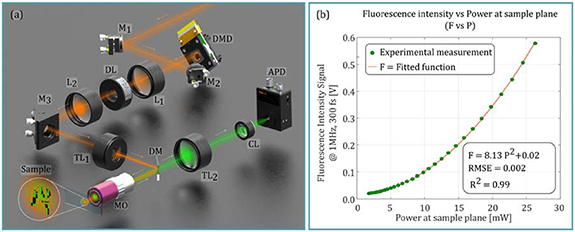
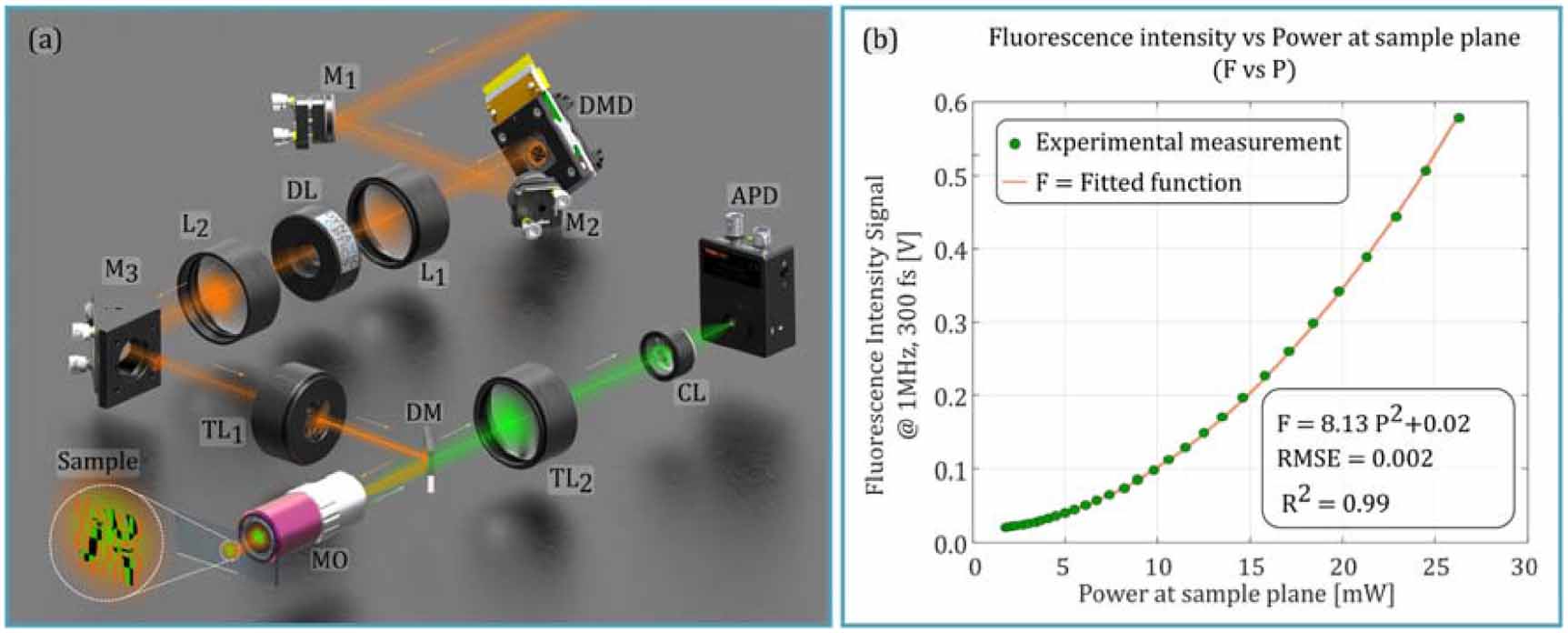
2.1. Platform overview, operating conditions and non-linearity verificationA widefield-excitation 2P-SPM platform, hereafter denoted as 2P-SPM, was implemented by adapting established single-pixel microscopy building procedures and reconstruction principles [37, 38]. The 2P-SPM was built following the practical guidelines reported by Zapata-Valencia et al in [39, 40]. Excitation patterns were generated with a DMD, and the emitted fluorescence was collected with an APD Thorlabs bucket detector with reference APD410A/M. A render schematic of the optical layout is provided in figure 1 panel (a), while a complete description of the optical design, synchronization, and reconstruction pipeline is reported in the Methods section. The platform was intentionally operated in a regime that differs from most scanless widefield two-photon implementations [20–22, 24]. In our approach, the cost-effective ultrafast sources and conventional optics are combined with scalable single-pixel techniques without relaying in complex TF, to broaden access to widefield-excitation 2PM. From now, the widefield-excitation term will denote simultaneous illumination of an extended sample region by each projected pattern, rather than point-by-point raster excitation; it does not denote a field of view larger than that achievable with galvanometric scanning.
Figure 1. 2P-SPM experimental setup and non-linear evaluation. In panel (a) a detailed render schematic of the optical set-up is provided. In panel (b) the recovered signal nonlinearity variation corresponding to the excitation produced by one pattern while the laser power is increased is shown. The detailed element in the schematics corresponds to Mi: mirror ith, Li: lens ith, DL: Deformable lens, TLi: tube lens ith, DM: dichroic mirror, MO: microscope objective, CL: condenser lens, APD: avalanche photodiode bucket detector APD410A/M.
Download figure:
Standard image High-resolution imageExcitation was provided by an industrial-grade Coherent Monaco laser at 1064 nm delivering 300 fs pulses, and imaging was performed without TF. In contrast to temporally focused widefield architectures, temporal pulse re-compression at the sample plane is intentionally omitted, and the excitation path is not engineered to relay a spectrally dispersed diffraction cone through the objective pupil [20, 22, 27] .As a result, the system optical architecture can be kept compact and alignment-tolerant, and the dispersive relays and tight dispersion-management requirements associated with TF modules are avoided [27, 28].
In addition to the robust and simple optical system, versatility at the sample plane is also achieved. The optical system was built to remain compatible with standard microscope components and to preserve comfortable mechanical access to the specimen. A 20×/0.4-NA objective was employed, and a working distance of approximately 20 mm was maintained. This working distance is central to the proposed use case, because it enables handling of a broad range of optical samples and non-standard holders, including thick substrates and custom fixtures, while avoiding the mechanical constraints imposed by the typically short-working-distance, high-NA objectives [41].
Operating without TF and without specialized dispersion pre-compensation requires that the nonlinear signal generation be verified under patterned excitation before further performance claims are made. For this reason, a thin fluorescent dye layer deposited on a microscope slide was used as an initial reference specimen. One spatially coded excitation pattern was projected over the sample, as seen in the zoom-in of the sample in figure 1 panel (a), and the integrated fluorescence was recorded with the bucket detector as a function of the power at the sample plane. In figure 1 panel (b) the clear nonlinear dependence of the detected signal on excitation power was observed. This behaviour confirms that robust two-photon emission can be generated and measured under the proposed operating conditions. This validation establishes the premise of the work: despite the deliberate removal of TF and the use of standard optics with long working distance, reliable nonlinear measurements can be obtained when excitation is pattern-coded, enabling subsequent evaluation of depth-dependent contrast and reconstruction performance in the following sections.
2.2. Widefield-excitation Hadamard-based SPIHaving validated robust two-photon emission under patterned excitation in the absence of TF, the next step is to demonstrate that the measured bucket signal supports stable single-pixel image formation. Among the available sampling strategies for single-pixel microscopy, deterministic orthonormal bases such as Hadamard and Fourier are widely used. In low-photon and noise-limited conditions, Hadamard sampling is often preferred due to the noise robustness while it yields well-conditioned reconstructions, particularly when implemented with complementary pattern pairs for differential measurements [33].
In Hadamard single-pixel microscopy (HSPM), a sequence of binary patterns  is sequentially demagnified onto the sample
  . For each pattern, the total emitted fluorescence is integrated by a bucket detector, generating a measurement vector
  [29, 32, 39]. Each measurement can be expressed as an inner product between the sample and the illumination pattern,

where  is the magnification ratio, and
  accounts for noise and drift at each measurement. After acquisition of a complete basis, the image is recovered by the inverse Hadamard transformation. When only a subset of patterns is acquired, a regularized inversion is performed, as detailed below and formalized in the methods section.
In figures 2, 64 × 64 pixel images were obtained for the initial demonstrations. Under this condition 4096 measurements are required at full sampling and 2048 at a sampling ratio (SR) of 50%. At the DMD refresh rate used here, these values correspond to nominal pattern-display times of 0.14 s and 0.07 s, respectively, or twice these values when complementary binary patterns are used. The complete experimental acquisition times were 0.16 s and 0.09 s, respectively, including detector integration, synchronization, and data transfer. The effective sampling pitch at the sample plane is set by the DMD pixel pitch, the chosen binning factor, and the system magnification. These parameters were selected to ensure reliable pattern transfer over the field of view. Full details of pattern encoding, binning, projection calibration, and timing are provided in the Methods section. For reference using a conventional high-speed resonant galvo scanner of 12 kHz, for the same number of pixels, would result in an imaging time of 0.34 s.
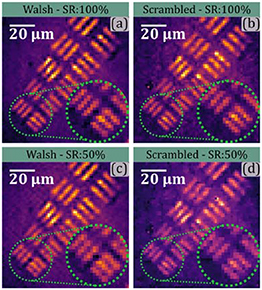
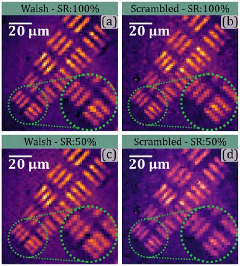
Figure 2. Widefield-excitation two-photon Hadamard single-pixel imaging and compressive reconstruction. In panels (a) and (b) the Walsh and the Scrambled reconstruction of the Group 7 of an USAF test target covered by a thin layer of fluorescent marker are shown. In panel (c) and (d) the corresponding reconstructions using only 50% of the sampling patterns is shown. For the Walsh Hadamard approach a ZigZag/TV sampling was implemented while for the scrambled case a random approach was employed. A resolving capability of approximately 2.1 µm is validated for both full sampling and compressive approaches.
Download figure:
Standard image High-resolution imageFigure 2 panels (a) and (b) present the two-photon single-pixel reconstructions obtained with the platform on a fluorescent stained USAF resolution target. Two acquisition variants were evaluated. First, a conventional ordered Walsh-ordered sequence was used as a baseline. The resulting image is shown in figure 2 panel (a). Second, a scrambled block Walsh–Hadamard strategy was employed to redistribute the spatial-frequency content across the measurement set. The construction of the scrambled Hadamard sampling base is detailed in the methods section and analysed in the supplementary note 1. The image reconstructed with the scrambled strategy is shown in figure 2 panel (b) [42, 43]. Stable image formation is obtained in both cases; the imaged Group 7 elements confirm that the target resolution is preserved. This corresponds to an achievable resolution of to 2.1 µm. The scrambled basis is of particular interest in the present non-temporal-focusing regime, as it reduces the dominance of low-order, slowly varying modes [44]. This aspect becomes central in the next section, where it is shown to influence the depth-dependent response of patterned excitation and, consequently, the achievable partial sectioning.
To reduce acquisition time and demonstrate measurement efficiency, CS was then applied. A 50% SR was used as an initial operating point, and reconstructions were computed using a NESTA-based regularized solver [45, 46]. In that way it is possible to reduce the number of patterns to retrieve the final image. Therefore, under this imaging conditions the 2P-SPM approach outperforms traditional point by point scanning since it is not necessary to measure the complete NxN image point. The optimization problem, solver settings and download links are provided in the supplementary note 2. As shown in figure 2 panel (c) and (d), comparable structural content is retained under compression, supporting compressive acquisitions as a practical operating mode for two-photon SPI. Being able to resolve details up to 2.1 µm for both the full sampling and the compressive approach.
2.3. Partial sectioning via propagation of structured excitationOnce robust two-photon single-pixel image formation on a resolution-standard target has been stablished, we characterize the axial response of the platform, and determine whether depth-dependent contrast can be achieved without TF. In this work, the term partial sectioning denotes a practical, depth-selective response in which out-of-plane contributions are progressively suppressed over a finite axial range. This behaviour differs from temporal-focusing-based widefield confinement [20–22, 24] and from the intrinsic sectioning associated with point-scanning 2PM. Neither approach is implemented here. Instead, partial sectioning arises from the propagation-dependent loss of pattern modulation when structured excitation is projected away from the DMD conjugate plane.
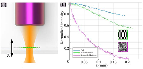
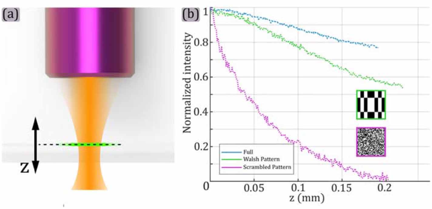
To demonstrate this effect, a thin fluorescent dye layer on a microscope slide was axially translated while the integrated fluorescence was recorded for a single projected pattern. All measurements were performed under fixed excitation conditions, the Monaco Laser was set to 1 MHz repetition rate, 300 fs pulse width, and a constant average power of 20 mW at the sample plane. The projected patterns were encoded on a 64 × 64grid with a binning factor of 3. Three illumination cases were compared: (i) a fully-on DMD state, equivalent to a ‘uniform’ widefield illumination, (ii) a representative Walsh-ordered Hadamard pattern, and (iii) a representative scrambled Hadamard pattern.
In figure 3 the normalized bucket fluorescence of the thin fluorescent layer as a function of axial displacement z is shown for different illumination patterns. Under uniform illumination, with a completely white pattern encoded on the DMD, the detected signal decreased slowly with the axial displacement. This is consistent with the absence of TF and with the fact that a spatially uniform field carries no high-frequency modulation that can be degraded by defocus. In contrast, patterned excitation produced a markedly stronger depth dependence. As the sample is displaced away from the projection plane, diffraction and defocus progressively wash out the spatial modulation of the projected pattern. The effective pattern contrast at the specimen therefore decreases, and the bucket detector becomes less sensitive to spatial structure outside the intended plane. This modulation-loss mechanism provides a route to partial sectioning without pulse re-compression, high-NA pupil filling, or dedicated dispersion compensation.
Figure 3. Pattern-driven depth selectivity without temporal focusing. In panel (a) the schematic representation of the axial displacement of a thin fluorescent layer relative to the objective focal plane is shown. In panel (b) the normalized bucket fluorescence as a function of axial displacement is shown. Uniform excitation (DMD fully-on), Walsh-ordered Hadamard, and scrambled Hadamard patterns (all encoded on a 64 × 64 grid with binning factor 3) are shown. Fixed excitation conditions, 1064 nm, 1 MHz, 300 fs, P avg = 20 mW are employed in all cases.
Download figure:
Standard image High-resolution imageThe comparison between the conventional and scrambled patterns clarifies why scrambling becomes essential in the present non-temporal-focusing regime. Squared insets in figure 3(b) shows the analysed patterns for the Walsh-ordered Hadamard case and the scrambled Hadamard case. Walsh-ordered Hadamard sequences contain many low-order modes with slowly varying spatial structure. These components are comparatively propagation-robust and behave similarly a DC-like contribution in the bucket measurements. They sustain out-of-plane background and weaken depth selectivity. Scrambling redistributes spatial-frequency content across the projected patterns and reduces the influence of these propagation-robust components. Consistent with this interpretation, the scrambled pattern exhibits a stronger attenuation with axial displacement, as is shown in the plot in figure 3(a).
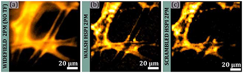
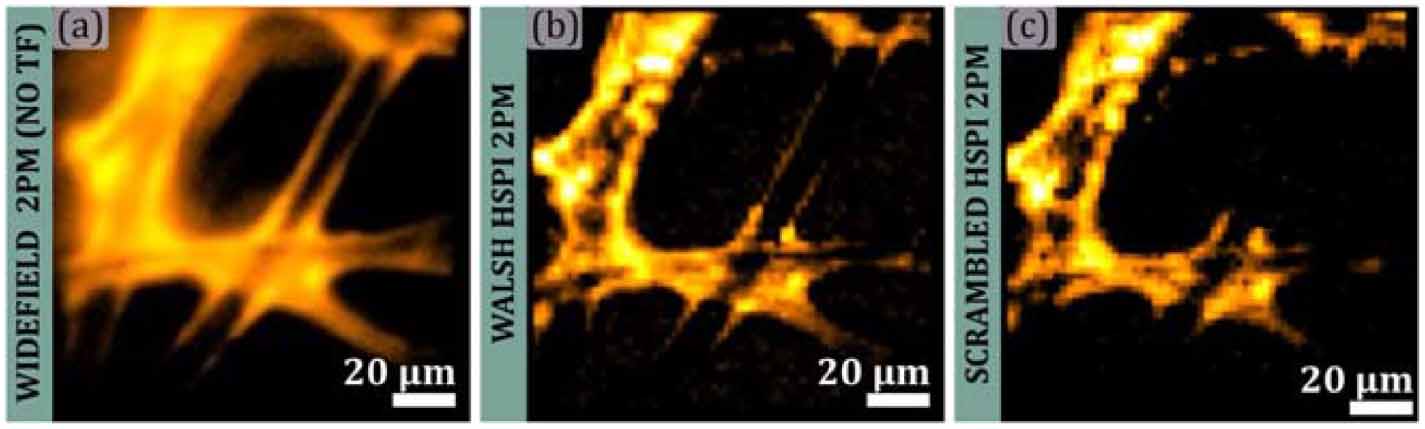
Representative two-photon fluorescence images further illustrating the central finding of this work are presented in figure 4. Under conventional widefield excitation, fluorescence contributions from both the focal plane and out-of-focus regions are simultaneously detected, producing a diffuse background across the image. These out-of-focus contributions remain visible in the Walsh-ordered reconstruction of the cotton tissue, although the principal tissue structures are preserved. In contrast, scrambled Hadamard sampling substantially reduces these contributions while retaining the main in-focus features, thereby providing partial depth-dependent rejection without TF.
Figure 4. Two-photon fluorescence imaging of cotton tissue using widefield and single-pixel acquisition. (a) Widefield two-photon fluorescence image acquired without temporal focusing. (b) Hadamard single-pixel image reconstructed using conventional Walsh sampling. (c) Hadamard single-pixel image reconstructed using the proposed scrambled sampling strategy. The scrambled approach reduces background and out-of-focus contributions while preserving the principal tissue structures. Scale bars: 20 µm.
Download figure:
Standard image High-resolution imageTwo implications follow from these results. First, depth-dependent contrast can be recovered in a widefield-excitation two-photon single-pixel architecture without TF, even when using moderate-NA and long-working-distance optics. Second, the observed axial selectivity is not incidental; it is an engineerable consequence of pattern propagation and basis design. This observation identifies pattern design as a practical control knob for depth-dependent contrast in nonlinear single-pixel microscopy, motivating systematic exploration of propagation-aware bases beyond conventional Walsh ordering. The practical impact of this choice is illustrated by the representative reconstructions in figures 4(b) and (c), where scrambled sampling suppresses diffuse out-of-plane contributions relative to Walsh ordering under comparable acquisition conditions. On this basis, scrambled Hadamard sampling is adopted for the subsequent experiments. Section 2.4 then tests whether the same mechanism yields improved feature separability in a weakly turbid biological specimen, where camera-based widefield acquisition without TF is expected to remain background-limited.
2.4. 2P-HSPM biological validationThe practical relevance of the proposed operating regime was next assessed on a biological specimen. A stained stoma from a cactus leaf was selected as a representative weakly turbid sample, where camera-based widefield detection is expected to be affected by diffuse background and by the loss of spatial information carried by multiply scattered emission. This experiment therefore evaluates whether the depth-dependent contrast observed under patterned excitation translates into improved feature separability in practice, while maintaining the low-complexity optical configuration and moderate-NA, long-working-distance imaging conditions.
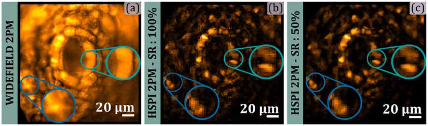
Figure 5 panel (a) shows a camera-based widefield two-photon image acquired without TF under uniform excitation with the DMD fully on. For this measurement, a 128 × 128 fully-on pattern was displayed on the DMD using the same binning factor of 3 adopted throughout this section, resulting in an effective imaging field of approximately 200 × 200 µm at the sample plane. The bucket detector was replaced by a camera to provide a direct widefield reference under identical excitation conditions. As expected, for scanless widefield excitation in this non-temporal-focusing regime, a strong diffuse background is observed and local features appear partially merged, consistent with the integration of out-of-plane and scattered fluorescence contributions.
Figure 5. Cactus stoma imaging: widefield 2PM versus proposed 2P-HSPM. In panel (a) a camera-based widefield two-photon image acquired without temporal focusing under uniform excitation (DMD fully-on) is shown. In panel (b) two-photon Hadamard single-pixel reconstruction using a 128 × 128 scrambled Hadamard basis (SR = 100%) is shown. In panel (c) a compressive reconstruction using a SR = 50% of the same sample is shown. Highlighted regions indicate improved feature separability relative to widefield acquisition. Scale bar, 20 µm.
Download figure:
Standard image High-resolution imageFigure 5 panel (b) reports the corresponding 2P-HSPM reconstruction acquired over the same 128 × 128 grid with binning factor 3, corresponding to a field of view of ∼200 × 200 µm. A scrambled Hadamard basis was used at SR = 100%. Compared with the widefield reference, the stomatal structure is more clearly delineated and localized emitters are more readily separable from the surrounding background. The highlighted regions mark representative locations where contrast is reduced in widefield detection, but structural detail is recovered through correlation-based single-pixel reconstruction.
Beyond biological validation, this experiment also demonstrates scalability to larger reconstructed grids while remaining within the same non-temporal-focusing operating regime and using the same industrial 1064-nm source. Increasing the reconstruction from 64 × 64–128 × 128 expands spatial coverage but increases the measurement count and therefore acquisition time. Figure 5 panel (c) shows a reconstruction computed at SR = 50% using the same scrambled Hadamard measurements, preserving the principal morphological features and maintaining the contrast improvement relative to widefield imaging while reducing the number of projected patterns by a factor of two.
Overall, the stoma experiment provides a direct biological validation of the operating regime introduced in this work. Improved feature separability is achieved through depth-dependent contrast in a widefield two-photon configuration without TF, without high-NA short-working-distance objectives, and without dedicated pulse-chirp control or elaborate dispersion compensation [12, 27]. At the same time, the 128 × 128 demonstrations establish a practical route to larger fields of view, while compressive acquisition provides a straightforward mechanism to manage the associated increase in measurement time. These results position pattern-coded, correlation-based two-photon SPI as a simplified and scalable alternative for widefield microscopy in weakly turbid specimens, where camera-based acquisition without TF remains background-limited.
This work establishes a distinct operating regime for widefield-excitation 2PM by demonstrating that two-photon SPI can be performed without TF, without sub-200-fs pulse management, and without high-NA, short-working-distance objectives. The relevance of the 300-fs source employed here does not arise from pulse duration alone, because multiphoton imaging has been demonstrated in substantially longer temporal regimes. Rather, its 1-MHz repetition rate provides sufficient pulse energy for extended-area patterned excitation without a temporal-focusing module. This simplification entails a trade-off: unlike TF, the present architecture does not intrinsically confine two-photon excitation to a thin axial plane. The observed depth-dependent contrast instead results from the propagation sensitivity of the projected patterns and should therefore be described as partial or propagation-dependent contrast, not as equivalent to the axial sectioning of TF. These results show that scanless multiphoton operation does not strictly require temporal pulse re-compression at the sample plane, nor the dispersive relays, pupil-filling constraints, and stringent alignment tolerances that commonly accompany TF-widefield implementations.
A second contribution is the identification and exploitation of a mechanism for depth-dependent contrast in the absence of TF. Under uniform widefield excitation, axial selectivity is not expected and camera-based detection remains background-limited by diffuse contributions. Here, partial sectioning arises from pattern propagation: structured excitation undergoes diffraction- and defocus-driven modulation loss away from the projection plane, reducing the sensitivity of bucket measurements to out-of-plane structure over a finite axial range. The results further show that this behaviour is governed by basis design. Walsh-ordered Hadamard sampling over-represents slowly varying low-order modes that remain propagation-robust and contribute a DC-like term to the bucket measurements, whereas scrambled Hadamard sampling redistributes spatial-frequency content and strengthens depth selectivity. This establishes a practical operating point between point-scanning 2PM and temporally focused widefield systems, retaining useful depth-dependent contrast while avoiding the optical burden typically associated with scanless multiphoton excitation.
The proposed architecture should be regarded as complementary to TF rather than as a universal replacement. TF remains advantageous when intrinsic axial confinement over an extended excitation area is the dominant requirement, although its implementation generally requires a sufficiently broadband ultrashort-pulse source, dispersive optics, accurate pupil relaying, and careful management of spatial and temporal dispersion. Strong confinement is also commonly achieved using relatively high-NA objectives, with the associated reductions in working distance and sample accessibility. By contrast, the present system prioritizes a simpler optical structure, compatibility with long-working-distance moderate-NA objectives, bucket detection, flexible basis engineering, and compressive acquisition. Avoiding the temporal-recompression stage reduces the number of specialized components and the corresponding alignment and cost requirements, providing an initial step toward more broadly accessible widefield-excitation 2PM. Shorter pulses with suitable bandwidth could increase nonlinear excitation efficiency and permit a hybrid temporal-focusing implementation, while a higher-NA objective could improve lateral resolution, excitation confinement, and fluorescence collection. These changes would, however, require renewed dispersion management and pupil relaying and would increase system complexity and cost while generally reducing working distance and mechanical accessibility. Moreover, higher NA alone would not make the observed propagation-dependent rejection equivalent to intrinsic temporal-focusing sectioning.
From an implementation perspective, the platform broadens access to widefield two-photon imaging in a manner that is directly relevant for experimental adoption. The use of an industrial-grade ultrafast source and conventional objectives, together with a long working distance on the order of 20 mm, supports integration with non-standard sample holders, microfluidic devices, and photonics fixtures where mechanical clearance and experimental flexibility are critical. Compatibility with compressive acquisition further provides a controlled trade-off between measurement count, acquisition time, and photon budget, enabling larger reconstructed fields of view without proportionally increasing acquisition time. While the depth-dependent contrast demonstrated here is not equivalent to the axial confinement achieved by TF or point-scanning 2PM, it defines a tunable design space for propagation-aware pattern engineering in nonlinear single-pixel microscopy and motivates future work on basis optimization, photon-efficiency improvements, and accelerated reconstruction for real-time operation.
4.1. Optical architecture and excitation sourceA widefield two-photon single-pixel microscope was implemented using structured excitation generated by a DMD and single-element fluorescence detection, bucket detection. Excitation was provided by an industrial femtosecond laser Coherent Monaco. The laser was set to operate at 1064 nm and 1 MHz repetition rate. The pulse duration was 300 fs. The system was operated without TF, and no dispersive grating or programmable pulse-shaping module was inserted in the excitation path. All the presented results were produced by following the guide reported guides for design, assembly, alignment and application of single-pixel microscope reported in [39]. All the required materials and an expanded protocol for the implemented HSPM construction can be found in repository contained in [40]. Then implemented DMD corresponds to a Vialux V-650 l DMD, with a pixel pitch of 10.8 µm. The structured light reflected by the DMD passes through the 2P-SPM setup until reaching the sample plane, where a demagnified image of the scanning patterns is projected. The microscope configures a telecentric imaging system with a Mitutoyo 20X microscope objective (MO), and a 200 mm Nikon tube lens (TL). The DMD is conjugated with both the object plane and the field diaphragm. A relay 4 F system, with lenses L1 and L2 of focal length 75 mm, offers direct access to the microscope aperture plane, which is directly conjugated with the pupil imaging system. In this plane a dynamic optics adaptive lens is placed to introduce an user define wavefront achieving the correction of the optical system induced aberrations [47]. The fluorescent sample light is collected by using a dichroic filter that ensures not residual IR is captured. A second TL2 and an aspheric condenser direct the light over the bucket detector.
4.2. Pattern projection and imaging optics fluorescence collectionBinary patterns were displayed on the DMD and optically relayed to the sample plane using a demagnifying telescope arrangement (TL and relay optics). Pattern grids of 64 × 64 and 128 × 128 were used, depending on the experiment. A binning factor of 3 was applied such that each effective pattern pixel corresponded to a 3 × 3 micromirror block. Two-photon excitation was delivered through a 20×/0.4 NA Mitutoyo microscope objective operated at an approximately 20 mm working distance. This long-working-distance configuration was selected to maintain compatibility with non-standard sample holders and to avoid the mechanical constraints associated with short-working-distance high-NA objectives. Epi-collected fluorescence was separated from the excitation beam using dichroic optics and emission filtering. The implemented dichroic mirror corresponds to a Thorlabs DMSP900R. For single-pixel acquisition, the emitted fluorescence was collected by an avalanche photodiode (APD; APD410A/M), used as a bucket detector. For each projected DMD pattern, the APD integrated fluorescence over the collection area and produced one scalar intensity measurement. The ordered set of measurements formed the intensity vector used for inverse Hadamard or compressive reconstruction. The APD therefore did not resolve the sample spatially; spatial information was encoded by the known illumination patterns. For widefield reference images, the bucket detector was replaced with a camera and the DMD was set to a fully-on state to approximate uniform widefield excitation without TF. In both cases an additional FESH0800 Thorlabs IR blocking filter was placed before the detection
4.3. Hadamard pattern sets and scrambling strategyHadamard single-pixel measurements were performed using two pattern orderings: (i) a conventional Walsh-ordered Hadamard sequence and (ii) a scrambled Hadamard ordering designed to redistribute spatial-frequency content across the pattern set and reduce the dominance of low-order, slowly varying modes. For full sampling (SR = 100%), the complete pattern set was acquired. The Matlab function fwht was implemented to perform the transforms and produce the imaging base. To produce the Scrambled based a random permutation was performed in the initial transform before the row reshaping. A detailed description of the algorithm and its implementation is provided in supplementary note 1.
4.4. Reconstruction: full sampling and compressive samplingCS represents a key point of the presented work. Since in conventional 2PM all the raster scanning must be projected in the proposed approach the projection time can be reduced by CS approaches. CS seeks to recover an image of the fluorescent sample  from
from  linear measurements by exploiting sparsity [48–51]. In the proposed the measured intensity vector
  are typically sparse in the Walsh Hadamard or the Scrambled Hadamard space, a necessary condition to exploit CS. In this sparse space is where we need to pick the relevant coefficients to be measured. Let
  denote the Walsh–Hadamard transform [36] and
  the row‐selection operator which is constructed by picking the most relevant coefficients the forward model to obtain our intensity vector
  measurements is described as follows:

Where
Comments (0)