Chemicals and reagents
Monocrotaline (MCT, 95%) was purchased from Aladdin Co. (Shanghai, China). Methanol, isopropanol, and formic acid (LC/MS grade) were from Macklin Biochemical Co., Ltd (Shanghai, China). Acetonitrile was from Adamas-beta (Shanghai, China). Neohesperidin, Hesperitin, Scopoletin, Esculetin, Neoeriocitrin, Lonicerin, Naringin, Naringenin, Limonin, Adenosine, Adenine, Syringic acid, Magnoloside A and Magnoloside B, Isosakuranetin, Xanthotoxol, Eriocitrin, Poncirin were from Sigma-Aldrich (Shanghai, China), Yangling Ciyuan Biotech Co., Ltd. (Shaanxi, China), Baoji Fang Sheng biological development Co., Ltd. (Shaanxi, China), Shanghai Sunny Biotech Co., Ltd. (Shanghai, China), Shanghai Standard Technology Co., Ltd. (Shanghai, China). The purity of these reference compounds were over 98%.
Preparation of ZXGD
Chinese herbal medicine in ZXGD were purchased from RenminTongtai Pharmacy (Harbin, China). According to the decocting method recorded in the Synopsis of Golden Chamber, 240g of Aurantii Fructus Immaturus and 240 g of Magnoliae Officinalis Cortex and 4000 ml distilled water were decocted to 1600 ml. After filtration, 480 g of Trichosanthis Fructus, 180 g of Allii Macrostemonis Bulbus, and 60 g of Cinnamomi Ramulus were then added into the filtration for another decocting and condensed into 1 g/ml. Finally, the decoction was filtered and freeze-dried.
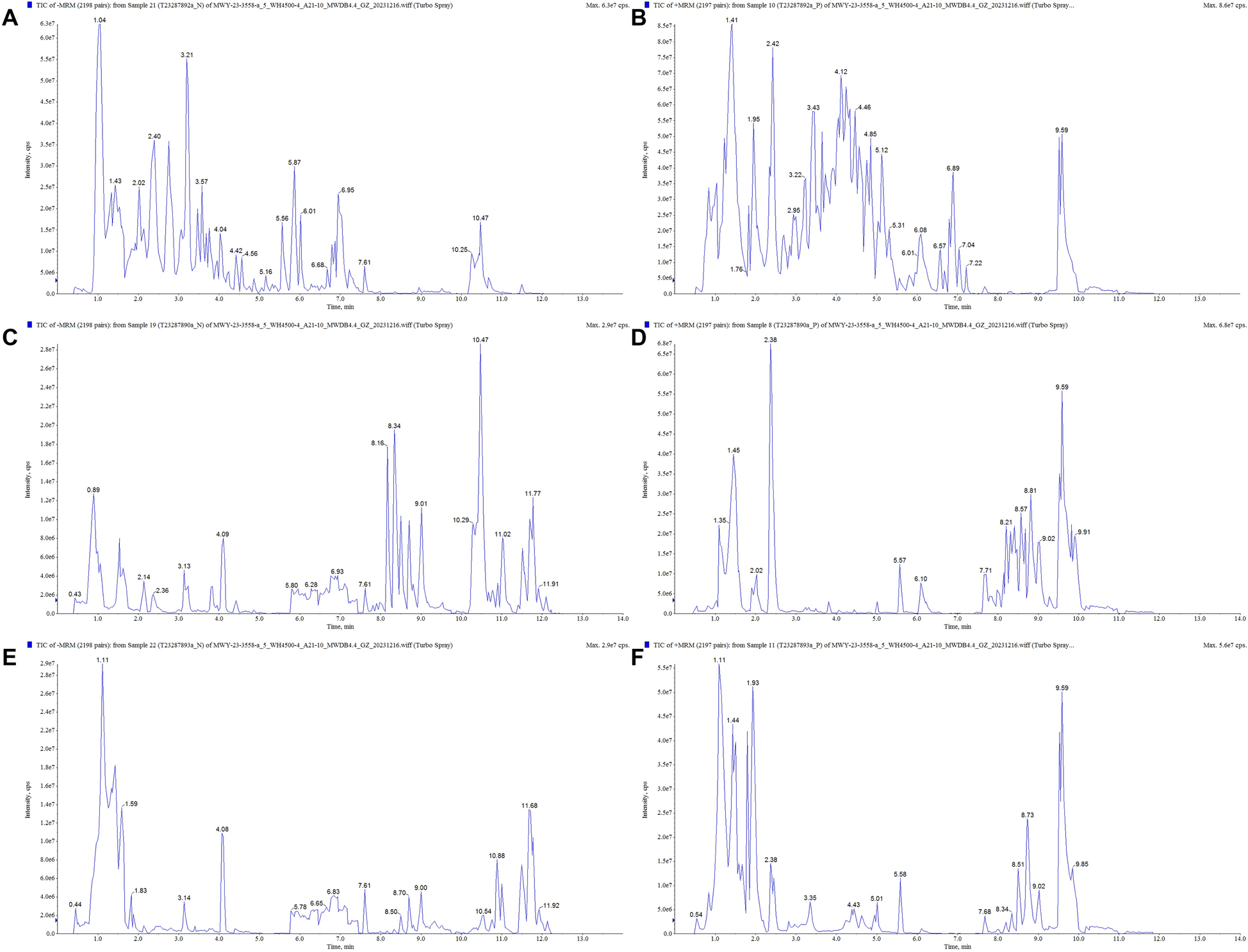
Chemical characterization of ZXGD
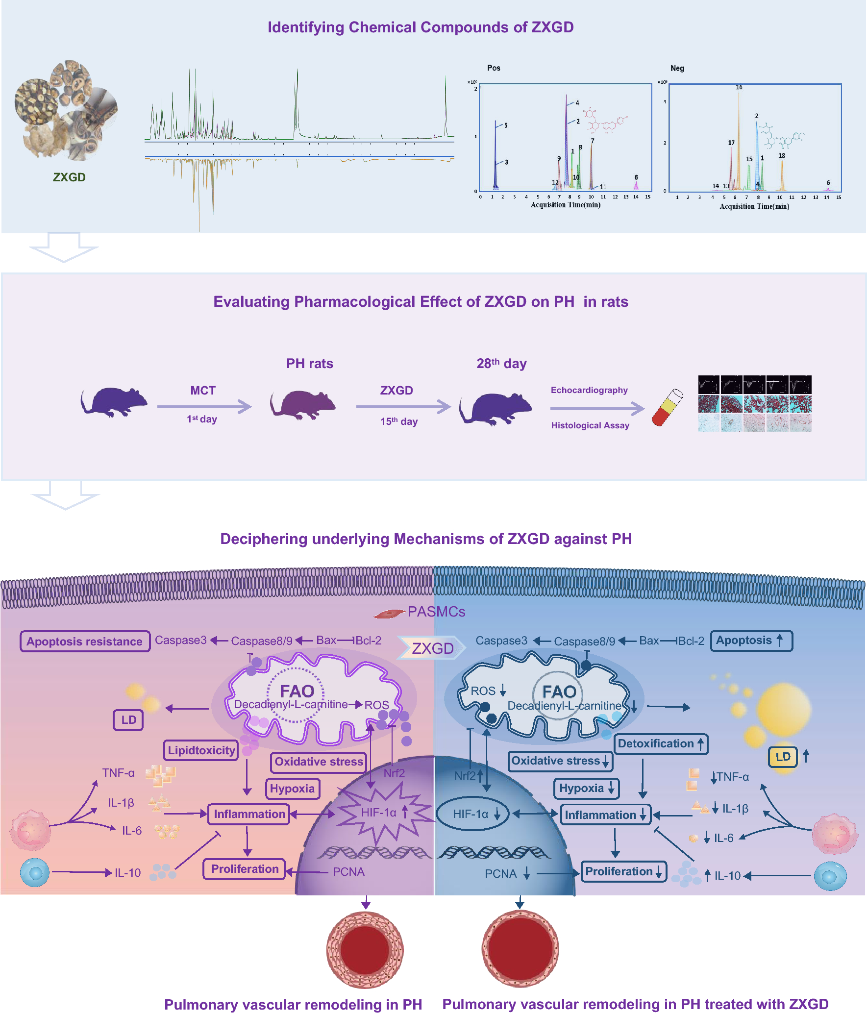
A UHPLC system (Agilent 1290 Infinity, Agilent Technologies, USA) integrated with QTOF-MS (Agilent 6560 IM-QTOF, Agilent Technologies, USA) were used to analyze the chemical compounds of ZXGD. An ACQUITY UPLC®BEH C18 column (2.1 × 100 mm, 1.7 μm, Waters) was applied for chromatographic separation of ZXGD. The mobile phase is composed of 0.1% formic acid with water (A) and 0.1% formic acid with acetonitrile (B). We provided the QTOF parameters in Supplementary Table 1. The information of 18 precisely identified compounds of ZXGD are listed in Supplementary Table 2.
Animals
Adult Sparague Dawley rats (male, weight of 200 ± 20 g) were obtained from the Experimental Animal Center of Harbin Medical University. All animal experiments methods in present study were carried out with the approval of the Ethical Committee of Laboratory Animals at Harbin Medical University (No. IRB3011621), Harbin, China, and with the conformity to the Chinese National Regulations on the Use of Experimental Animals.
We used PH rats induced with MCT because it is the most classical and widely used in vivo model for nearly 60 years due to its simplicity, reproducibility and low cost [42]. The following animal experiments were conducted: Rats were randomly assigned into five groups (n = 10, respectively), PH group, 2.7 g/kg ZXGD treatment group, 5.4 g/kg ZXGD treatment group, and 35 mg/kg Sildenafil treatment group rats were firstly intraperitoneally injected with 60 mg/kg MCT to induce PH occurrence for 28 days. Control group rats were received the same volume of saline as MCT. On the 15th day after MCT post-injection, rats in ZXGD treatment groups and Sildenafil group were orally administered with different dosage of ZXGD and 35 mg/kg Sildenafil daily for consecutive 14 days.
Echocardiography
All experimental rats were received a single intraperitoneal injection with chloral hydrated (300 mg/kg) to anesthetize to be prepared for echocardiographic examination. Echocardiographic assessment of pulmonary vascular function in rats was performed with a digital imaging platform coupled with linear array technology and Doppler mode (Vevo 2100, Visual Sonics, Toronto, Canada). Echocardiographic images were collected with pulsed wave Doppler, hemodynamic measurements of pulmonary blood flow including pulmonary arterial acceleration time (PAT), ejection time (ET), and PAT/ET were regarded as alternative indexes to estimate RVSP.
Histological assay
Using 4% paraformaldehyde, graduated alcohol and paraffin wax, the lung leaflets were received a fixation for overnight, then dehydration, subsequent embedding in paraffin wax. Further staining was performed with hematoxylin and eosin (H&E). The tissue pieces for immunohistochemistry were deparaffinized and rehydrated, then incubated with α smooth muscle actin (α-SMA) antibody (Boster, Wuhan, China) for overnight, subsequent 2 h incubation with goat anti-mice IgG. Brown color indicated positive stains. H&E and immunohistochemistry were visualized with an Eclipse 600 Nikon microscope and photographed with a digital camera, then analyzed using Image Pro Plus software. Measurement of diameter and area of each pulmonary microartery, then calculated wall thickness and positive stain area of α-SMA of pulmonary vascular.
Western blot
The methods in extraction, separation, and transferring membranes of protein samples from lung tissues and PASMCs were performed according to our previous study [24], further incubation with the following primary antibodies: Caspase 3, Caspase 9, PLA2, PPARγ, IL-1β, IL-10 (Bioss, Beijing, China); Bax, HIF-1α, PLIN2 (Boster, Wuhan, China); PCNA (Wanleibio, Shenyang, China); IL-6 (Beyotime, Shanghai, China); Caspase 8, Bcl-2, β-actin (ABclonal, Wuhan, China) for overnight at 4 °C and subsequently incubated with secondary antibodies (goat anti-rabbit IgG, Bioss, Beijing, China) for 1h at room temperature. Immunoblots were scanned with Odyssey clx. The relative expression of protein to the control β-actin was determined by densitometric analysis using Quantity one software.
Enzyme-linked immunosorbent assay and biochemical analysis
Rat ELISA kits (MEIMIAN, Yancheng, China) were used to test the levels of decadienyl-l-carnitine, IL-1β, IL-6, IL-10, and TNF-α in lung tissue. Biochemical assay kits were employed to examine the serum concentration of TC, LDL-C and HDL-C in rats (Nanjing Jiancheng Bioengineering Institute, China). Measurement of Optical density (O.D) and calculation of concentration of each sample are based on a Microplate reader (Biotek, USA) and standard curve linear regression equation.
Cell culture
Acquisition and culture of PASMCs have been reported in our previous study [43]. Platelet-derived growth factor (PDGF)-BB was used to mediate PASMCs proliferation in vitro [44]. Before each experiment, the cells were subjected to growth arrest for 24 h, then exposed to different dosage of ZXGD, neohesperidin and naringin treated with or without 40 ng/ml PDGF-BB according to the experimental requirements.
MTT assay
PASMCs were exposed to different dosage of ZXGD, neohesperidin, and naringin with or without PDGF-BB treatment for 24 h incubation at 37 °C, then subjected to 20% FBS-DMEM medium including MTT reagent (Beyotime, Shanghai, China) incubation for 4 h, and blocked with DMSO. Subsequently, the samples were examined using a Microplate reader at 490 nm.
Oil red O staining
According to different experimental requirements, PASMCs were treated with 40 ng/ml PDGF-BB, 5 mg/ml and 10 mg/ml ZXGD, then incubated for 24 h. Next, incubated with 60% isopropanol for 5 min, rinsed twice with distilled water, subsequently oil red O dyeing solution (Beyotime, Shanghai, China) was added for 20–30 min, then hematoxylin dyeing for 1 min. Finally, microscope and Image Pro Plus software were applied for observation, visualization and analysis of PASMCs.
Measurement of ROS
PASMCs were exposed to 40 ng/ml PDGF-BB and different dosage of ZXGD (5 mg/ml, 10 mg/ml) for 24 h, then DCFH-DA probe (10 μM) (ROS Assay Kit, Beyotime, Shanghai, China) for 30 min at 37 ℃. CytoFLEX flow cytometry (FCM, Beckman Coulter, USA) and FlowJo 10.8.1 software were applied in this study for the detection and analysis of ROS.
Transient transfection of HIF-1α
The primer sequence of HIF-1α siRNA (GenePharma, Shanghai, China) and negative control (NC) were used as follow: HIF-1α siRNA sense 5′-CGCAUUGAA GUUAGAGUCAATT-3′ and antisense 5′-UUGACU CUAACUUCAAUGCTT-3′; NC sense 5′-UUCUCCGAACGUGUCACGUTT-3′ and antisense 5′-ACGUGACACGUUCGGAGAATT-3′. PASMCs cells were transfected with HIF-1α siRNA containing GP-transfect-Mate according to the manufacture’s instructions.
qRT-PCR
Total RNA was extracted from lung tissues and PASMCs with Trizol reagent (Beyotime Biotechnology, China) and reverse-transcribed using BeyoRT™II First Strand cDNA Synthesis Kit (Beyotime, Shanghai, China). The primer sequences of HIF-1α and β-actin were used as follows: HIF-1α forward 5′ TGGACTTGCCCC TTTCTCTG-3′ and reverse 5′-CGACGTTCGGAACTCATCCT-3; β-actin forward 5′-AAGTTCTACAAATGTGGC TGAGGA-3′ and reverse 5′-TCCTCTTAGGAGTGGG GGTGG-3′. Then, PCR was conducted using SYBR Green (Thermo, USA). The relative level of HIF-1α was calculated by the comparative Cq method (2−ΔΔCT).
Statistical analysis
One-way analysis of variance (ANOVA) is performed for statistical analysis using GraphPad Prism 5.0. All values were presented as mean ± SEM. p < 0.05 was set as statistical significance.


Comments (0)