Sex as a biological variable
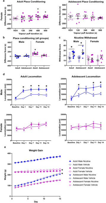
Both male and female mice (balanced for sex) were used in our study for all the behavioral and biochemical experiments with similar findings reported for both sexes (Supplementary Fig. 1). Therefore, in this study, sex was not considered a biological variable.
Animals
Med23flox/flox mice with Med23 exons 5–7 allele flanked by LoxP sites were described in our previous work [23]. By crossing Emx1-Cre mice [24] with Med23flox/flox mice, we generated Med23 CKO (Emx1-Cre:Med23flox/flox) mice for research. Littermates of other genotypes (i.e., Med23flox/+, Med23flox/flox, Emx1-Cre and wild type) were all used as controls. The mouse strains used in this study were kept in specific pathogen–free (SPF) conditions, and maintained on C57BL/6 J background.
Tissue preparation
Mice received a deep anesthesia with sodium pentobarbital dissolved in saline (40 mg/kg body weight) and then intracardially perfused with 4% paraformaldehyde (PFA) in phosphate-buffered saline (PBS). Brains were removed, postfixed in 4% PFA at 4 °C overnight, cryoprotected in 25% sucrose in PBS overnight and cut into 25 μm-thick coronal sections.
Nissl staining and immunohistochemistry
For Nissl staining, the sections were washed in PBS, stained with 1% crystal violet, followed by incubation in 95% and 100% ethanol, and xylene. Finally, the sections were mounted in neutral balsam with coverslips.
For immunostaining, the sections were incubated in antigen retrieval buffers (10 mM sodium citrate, 0.05% tween-20, pH 6.0) at 95 °C for 15 min. Then the sections were transferred into blocking serum (1% fetal bovine serum, 0.5% Triton X-100) for 1 h at room temperature and incubated with primary antibodies at 4 °C overnight. After being washed in PBS, the sections were stained with secondary antibodies (Alexa488-conjugated or Biotin-conjugated) at room temperature for 2 h. For those stained with biotin-conjugated secondary antibodies, 1-h incubation of cy3-conjugated streptavidin was applied. All slices were counterstained with Hoechst 33258 (Sigma, 94403, 1:2000) and then mounted in 75% glycerol with coverslips. The following primary antibodies were listed: anti-Ctip2 (Abcam, AB18465, 1:500).
Golgi staining
Golgi staining was carried out with FD Rapid GolgiStain Kit (PK401, FD NeuroTechnologies, USA) according to the manufacturer’s protocols. Solutions A and B needed to be mixed 24 h before use. PFA-fixed brains were placed in the premixed solution and refreshed the solution on the next day. After 2 weeks of incubation in the darkness, brains were transferred into solution C for 2–7 days, and finally were sectioned into 100-μm-thick slices. The secondary dendritic branches of the granule cells in the DG were selected to analyze spine density and the proportion of different categories with four spine shapes.
Western blot
The hippocampal tissues were lysed in precooled RIPA buffer containing 1% Triton-X 100, and protease inhibitor cocktails (ab2011111, abcam, UK). Samples were then boiled at 100 °C for 5 min to prevent protein denaturation. After cooling to room temperature, samples were loaded on SDS-PAGE and then transferred to a membrane filter, followed by incubating with 5% non-fat milk in TBST for 2 h at room temperature. After rinsing once with TBST, primary antibodies were applied overnight at 4 °C. All the antibodies were visualized by an ECL kit (34578, Thermo Fisher Scientific, USA), following incubating with HRP-conjugated anti-rabbit IgG (Proteintech, SA00001-2, 1:2000) for 2 h at room temperature. Primary antibodies used for western blot in this study were listed: anti-DRIP130/Med23 (Abcam, ab200351, 1:1000) and anti-GAPDH (Proteintech, 81640-5-RR, 1:3000).
RT-qPCR
Total RNA was extracted from the hippocampus with RNAiso Plus following the manufacture’s protocol (9109, TaKaRa Biotechnology, Japan) and then the PrimeScript™ RT reagent Kit (TakaRa_RR037A, TaKaRa Biotechnology, Japan) was used to generate cDNA. Each sample was triplicated in RT-qPCR using ABI-Q7 (Applied Biosystems, USA) with RT² SYBR Green ROX qPCR Mastermix (330524, Qiagen, Germany). The sequences of primers are listed:
Med23-F:3ʹ-TCGGAAAATCATTGGAGGAG-5ʹ;
Med23-R:3ʹ-CAATAGGCAGGCATTTCGTT-5ʹ;
GAPDH-F:3ʹ-AACTTTGGCATTGTGGAAGG-5ʹ;
GAPDH-R:3ʹ-ACACATTGGGGGTAGGAACA-5ʹ.
Behavioral tests
Adult (3–6 months old) mice were used in the following behavioral tests. Behavioral experiments were all performed during the light phase of the 12 h light/dark cycle, and behavioral tests were carried out in sound-proof rooms with a neutral environment. Mice were transported to the experiment room 30 min beforehand for habituation, and behavioral tests included the open field test, object-based attention test, elevated plus maze, Y maze, cliff avoidance reaction (CAR) and pre-pulse inhibition test. The experimenter was blind to the genotypes of mice in the tests.
Open field test
The open field apparatus comprised a square arena with walls around that are all made of white acrylic plates (40 × 40 × 40 cm, L × W × H). The activities of mice in the apparatus were captured and analyzed automatically by computer (Omnitech SuperFlex, Omnitech Electronics Inc, USA). The center square of 20 × 20 cm was set as the center zone while the other area was the peripheral zone. Mice were allowed to explore the apparatus freely for 30 min. Total traveling distance, distance traveling per 10 min, ambulatory time, average velocity, times spent in center or peripheral zone and vertical or stereotypic activity were recorded.
Y maze
Spontaneous alternation behavior (SAB) in the Y maze test requires attention and working memory [25]. During the tests, mice were placed individually at the end of an arm and allowed to explore the maze freely for 10 min. The order of arm entry and total number of arm entries were recorded manually. An arm entry was defined as the center of the body into one arm. The actual alternations (successive entries into the 3 different arms of the Y-maze without overlapping) and the maximum number of alternations (the total number of arm entries minus 2) were calculated. The SAB score was the ratio of (the actual alternations/ the maximum number of alternations).
Object-based attention test
The procedures for object-based attention test were adapted from the study previously described to fit the facilities [26]. Mice were placed in empty chambers (the exploration chamber and the test chamber, 33 × 33 × 40 cm, L × W × H) each for 10 min on day 1–2 for habituation. On day 3 for the test phase, five objects (A, B, C, D, E) of the same size but different shapes (square-, star-, heart-, hexagon-, and cross-shaped) were placed in the chamber. Mice were allowed to explore freely in the chamber for 5 min during the exploration session and then they were transferred to a test chamber containing a familiar (square-shaped, object A) and a new (round-shaped, object F) object in less than 10 seconds and allowed to explore these two objects for a 5 min of retention session. Throughout the study, the position of each object in the exploration and test chambers was fixed. The recognition index (RI), calculated for each mouse, was expressed as the ratio of (TF × 100)/(TA + TF), where TA and TF are the time spent on object A and object F during the retention session, respectively. Another recognition index calculated in this test was expressed as the ratio of (TA × 100)/(TA + TD), where TA and TD are the time spent on object A and object D during the exploration session, respectively. Noldus software (EthoVision XT 15.0, Noldus Technology, Netherland) was used to monitor and track the movement of mice.
Cliff avoidance reaction (CAR)
The cliff avoidance reaction (CAR) is used to evaluate maladaptive impulsive rodent behavior, especially for ADHD animal models [27]. The apparatus included a round wooden platform (20 cm in diameter and 2 cm in thickness) and a heavy iron rod (50 cm in height) below. The test began with placing mice on the platform with the forelimbs near the edge of the platform and lasted for 30 min. Falling from the platform once was recorded as one impaired CAR. After falling down, mice were placed back on the platform immediately. The distance traveled on the platform in the 30 min and the latency from the beginning until falling and the number of falls were recorded with Noldus software. The incidence of impaired CAR was calculated as a percentage index for each group, as follows: % CAR = (the number of mice that stay on the platform/the total number of mice) × 100.
Differential reinforcement of low rate schedule (DLR)
The procedures for DLR were adapted from the study previously described to fit the facilities [28, 29]. The apparatuses used were 4 operant chambers, each containing a touchscreen box (Campden Instruments, 89540 A). Briefly, the touchscreen box was equipped with 2 nose poke touchscreens, a loudspeaker, a liquid dispenser and a liquid tray with a LED light above it. Mice were weighed and handled for 1 min in the first 3 days and then underwent a food deprivation until their body weight reduced to 85–90% of their free-feeding weight. Strawberry milkshake was provided to the mice in their home cages for 2 days immediately before the training phase. The training phase can be divided into 3 stages under a continuous reinforcement schedule.
In the first stage, food-deprived mice were first trained to approach the tray after an LED light and an auditory stimulus to obtain a strawberry milkshake reward (10 μL). In the second stage, mice are trained to nosepoke the touchscreen, where an image pseudo-randomly appears on one of the two screens. Mice nosepoking the screen without an image received a strawberry milkshake reward (10 μL), while nosepoking the screen with an image resulted in a five times greater reward (50 μL). In the third stage, mice nosepoking the screen with an image received a strawberry milkshake reward (10 μL), while nosepoking the screen without an image received no reward. All the training stages lasted for at least 3 daily sessions of 30 min, or until a performance criterion (>30 rewarded responses within a session) was reached.
After the training phase, the DLR task was conducted. In this task mice were required to wait for a period of time (inter-response-time, IRT) between 2 consecutive nosepoke responses. Premature nosepoke reset the delay and resulted in no reward. Mice were trained and tested successively on DRL-4 s, DRL-6 s, DRL-8 s, DRL-10 s, DRL-15 s, each for 3 daily sessions of 30 min (30 trials within a session) and the result of the final session was analysed. The ratio between reinforcements and the total number of responses were calculated for DRL-4 s, DRL-6 s, DRL-8 s, DRL-10 s and DRL-15 s. Responses with IRT < 1 s were regarded as bursting response, while responses with IRT > 30 s were regarded as long IRT. The number of bursting responses and long IRT were calculated for DRL-15 s. For qualitative assessment of timing behavior, the relative frequency distributions of IRTs were generated, using a 5 s bin size.
Pre-pulse inhibition test (PPI)
Experiments were performed in sound attenuating test chambers (33 × 35 × 48 cm, L × W × H) equipped with a commercial startle reflex system in each chamber (SR-LAB Startle Response, San Diego Instruments, USA). First 5 min of the test is the acclimation phase with 50 dB acoustic background noise. Then 120 dB startle pulse (20 ms) was given to mice 12 times. In 48 subsequent experiments, the scare sound appeared either alone or after a random delay of 100 ms at three pre-pulse intensifies (65, 72, and 83 dB, 20 ms). The average interval between each trial was 30 s (random number from 20 to 40). The average startle amplitude during the 100 ms following the onset of each startle stimulus was recorded automatically. The first 12 trials are used to measure the baseline of acoustic startle response but excluded in the PPI analysis. The amount of PPI was expressed as the percentage decrease in the amplitude of the startle reactivity caused by presentation of the pre-pulse, which was calculated as follows: % PPI = 100 - .
Drug administration
Methylphenidate (MPH) is a widely-used in the treatment of patients with ADHD [30]. MPH effects in a dose-dependent manner and high-dose of MPH can cause adverse effects, such as anxiety and aggressivity [31]. The dose of 2.5 mg/kg is reported to be suitable according to previous works [32]. To understand the mechanism underlying the effect of MPH treatment on short-term synaptic plasticity in Med23-deficient granule cells, the noncompetitive N-methyl-d-aspartate (NMDA) receptor antagonist dizocilpine-MK801(M107, Sigma, USA) was applied [33]. Mice received a single intraperitoneal injection of MK801 with the dose of 1 mg/kg 30 min before MPH treatment. Both MPH (0.1 mg/ml) and MK801 (0.1 mg/ml) were dissolved in saline and these compounds were prepared on the day of the experiments.
In vitro patch-clamp recording
Hippocampal slices were prepared from mice by using procedures described previously [34]. The mouse brain was quickly removed and placed in ACSF bubbled with saturated gas (95% oxygen and 5% carbon dioxide) and cut into 350-μm-thick slices using a vibratome (Leica VT1000S, Leica Microsystems, Germany). All recording slices were carried out in the recording chamber, maintained at RT with standard ACSF, and placed on the Nikon microscope stage (600-FN, Nikon, Japan). The stimulation electrode was a pair of Teflon-coated 90 platinum/10% iridium wires. The recording electrode (1–3 mΩ) was pulled from borosilicate glass capillaries (1.5 mm outer diameter, 0.84 mm inner diameter, World Precision Instruments) with a Brown-Flaming micropipette puller (P-97; Sutter Instruments Company, USA), filled with ACSF. The fEPSP in the perforant path extending from layer II of the entorhinal cortex (EC) was recorded in the molecular layer of the DG area of the hippocampus by stimulation (0.1 ms duration). As a comparison, the fEPSP in the SC-CA1 pathway was recorded in the stratum radiatum of the dorsal CA1 area of the hippocampus by stimulation of the Schaffer collaterals (SCs) [35]. The GABA-dependent inhibitory transmission was not blocked during fEPSP recording. For input-output recordings, fEPSP slopes were recorded by increasing the intensity of stimulation (0.1 ms pulse width) from 0 with 20 µA increments. For paired-pulse ratio (PPR), the stimulation intensity was adjusted to give an fEPSP slope of 50% of the maximum, and a pair of stimuli with different intervals, including 50 ms, 100 ms, 150 ms, 200 ms, and 1000 ms was given successively for three times. The average value of three successive rounds was used for PPR analysis. The PPR was calculated as the ratio of the slope of the 2nd fEPSP to the slope of the 1st fEPSP. All data were recorded by using the pCLAMP 10 software (Molecular Devices). The signal collection was obtained by using Multiclamp 700B and Digidata 1440 A (Molecular Devices).
Statistical analysis
Statistical analyses were performed by GraphPad Prism 9.5.1 software (GraphPad Prism, USA). All values were expressed as the mean ± SEM. The number of samples indicates biological replicates and also are indicated with scattered dots in the bars in the figures. For analysis of spine morphology, more than 3 mice were included in each group. For behavioral experiments, over 8 mice are included in each group. Comparisons were made using Student’s t-test, multiple t-test, one-way or two-way ANOVA with Bonferroni correction as mentioned in each figure legend. Statistical significance was set at p < 0.05.
Study approval
Animal care practices and all experiments were reviewed and approved by the Animal Committee of Laboratory Animal Center, Fudan University, Shanghai, China (202408001Z).


Comments (0)