
Remember me
Palmar creases, the lines on the palms, provide valuable diagnostic clues for dermatological, genetic and systemic conditions, aiding in the recognition of both congenital and acquired disorders. This review examines their anatomical basis, variations, and disease associations, emphasising their importance in diagnosis and management while excluding the broader field of dermatoglyphics.
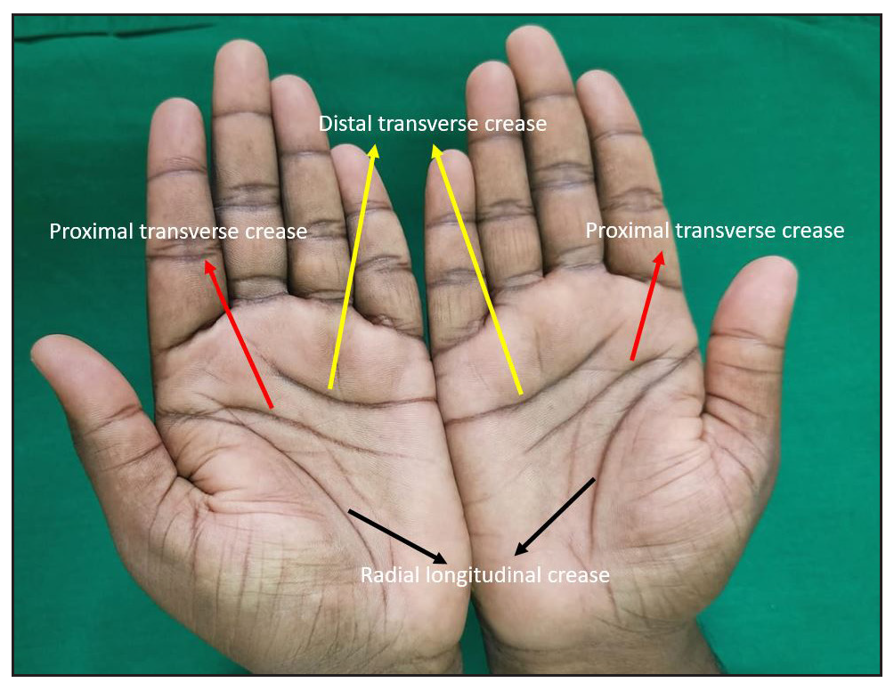
Types of palmar creasesPalmar creases are the epidermal flexure lines on the hand’s palmar surface, with three primary types: radial longitudinal crease (RLC), proximal transverse crease (PTC), and distal transverse crease (DTC) [Figure 1]. The radial longitudinal crease begins near or below the proximal transverse crease at the palm’s radial border, curves laterally, and extends toward the wrist. The proximal transverse crease starts at the radial side, runs medially with a slight curve, and ends at the hypothenar eminence. The distal transverse crease originates proximal to the index-middle finger interdigital space and extends toward the ulnar side, with a slight distal concavity. The proximal transverse crease and distal transverse crease typically do not traverse the palm’s full width. Palmar creases are classified based on one, two, or three radial origins.1 Variants include branched, forked, broken (detached segments), or cascade (closely running broken segments) patterns. Accessory creases, parallel to the main ones, may exceed half the length of the primary crease, offering significant diversity in palmar crease morphology and clinical relevance.2-4
Export to PPT
Abnormal palmar creasesPalmar creases are classified into Simian, Sydney, and Suwon creases based on the relationship between the proximal transverse crease and distal transverse crease. The Simian crease is a single transverse line seen in Down and Turner syndromes. The Sydney crease is an elongated proximal transverse crease crossing the palm, first described in Sydney. The Suwon crease, found in Suwon, Korea, spans the palm due to proximal transverse crease-distal transverse crease fusion or an extended distal transverse crease with an accessory PTC.5Aberrant palmar creases are observed in healthy individuals across various populations, including Nigerians, Koreans, Central Indians, and Americans. Their presence does not always indicate an underlying medical condition.6
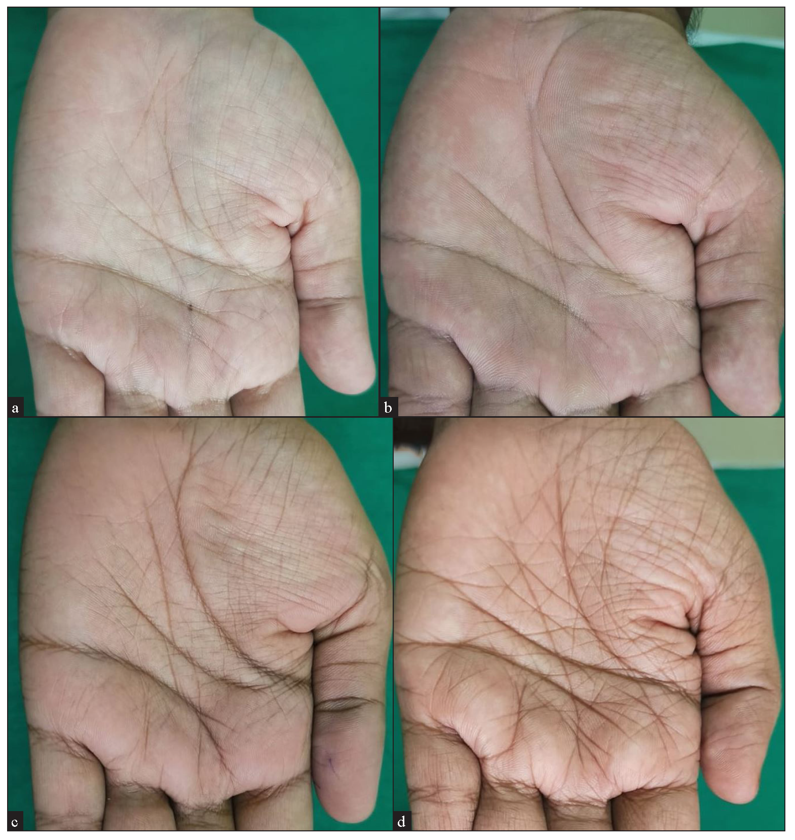
Clinical significance in dermatology 1. Hyperlinearity of palmsHyperlinear palms (HP) indicate abnormalities in keratinisation and are characterised by the presence of more than five distinct lines, each exceeding 1 cm in length, running across the palm, especially over the thenar eminence.7 The different patterns of palmar hyperlinearity can act as clinical markers for evaluating prognosis and treatment outcomes in atopic eczema. These patterns [Figure 2a-d] correspond to barrier function and disease severity, as summarised in Table 1.8 Notably, the extensive crosshatch pattern is closely linked to higher Eczema Area Severity Index (EASI) scores, Patient-Oriented Eczema Measure (POEM) scores, reduced skin hydration, increased transepidermal water loss and filaggrin (FLG) mutations. Finer patterns indicate milder disease and better barrier function, while crosshatch and diamond patterns signify more severe eczema and impaired barrier function.8
Export to PPT
Table 1: Patterns of Palmar hyperlinearity and their descriptions
Sr.no Types Pattern Description Significance 1 Pattern 1 Fine perpendicular lines Delicate, straight lines that run perpendicular to the palm’s length. Lower severity and better barrier function 2 Pattern 2 Fine crosshatch Subtle crosshatched lines that form a grid-like pattern. Lower severity and better barrier function 3 Pattern 3 Extensive crosshatch Dense crosshatching that covers a significant area of the palm.More severe disease and poorer barrier
function.
4 Pattern 4 Thick perpendicular Prominent, thicker lines that run vertically across the palm. Lower severity and better barrier function 5 Pattern 5 Prominent diamond Intersecting lines on the palm form distinct diamond shapes.More severe disease and poorer barrier
function.
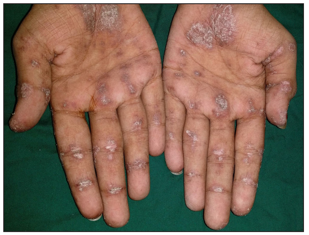
2. Inverse Gottron signThe inverse Gottron sign is a rare but specific cutaneous finding in dermatomyositis.9 It is characterised by painful erythematous papules and macules involving the palmar creases of the fingers [Figure 3]. It is a highly specific cutaneous marker of anti-MDA-5 positive dermatomyositis, linked to rapidly progressive interstitial pneumonia.10
Export to PPT
3. Kindler syndromeKindler syndrome is an autosomal recessive disorder characterised by acral blistering in infancy, photosensitivity, poikiloderma, cutaneous atrophy, and mucosal involvement. Common associations include periodontitis, gingivitis, palmoplantar keratoderma and diminution of palmar crease.11 In Kindler syndrome, palmar creases are notably diminished due to progressive cutaneous atrophy. This leads to a smooth, almost glossy appearance of the palms, with reduced dermal ridges and a loss of the typical fingerprint pattern.12
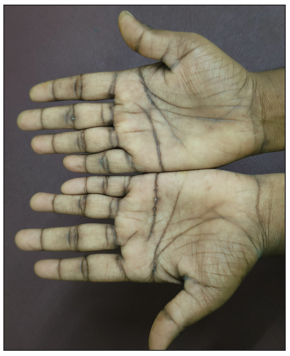
4. Keratosis punctata of the palmar crease (KPPC)KPPC is a benign dermatological condition primarily affecting African Americans (prevalence: 1.9–3.1%). It may be autosomal dominant or sporadic and is associated with Dupuytren contracture, knuckle pads, and striate keratoderma. It is characterised by 1–5 mm hyperkeratotic pits in palmar creases [Figure 4]; KPPC can cause discomfort, affect hand aesthetics, and hinder daily activities.13
Export to PPT
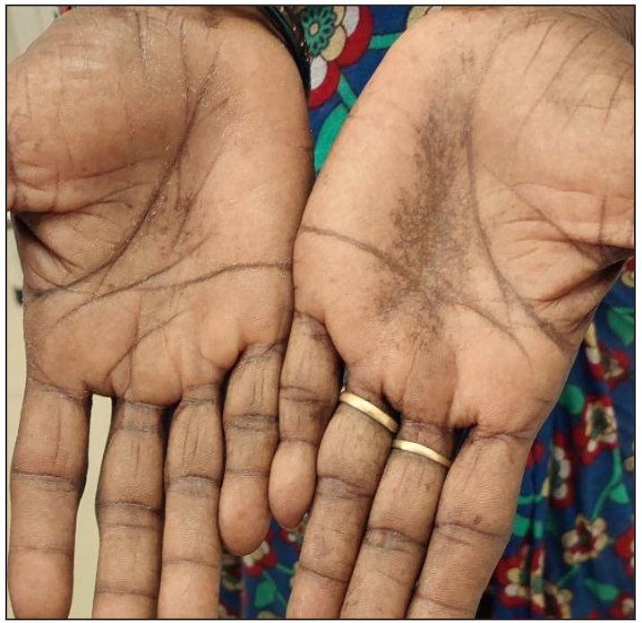
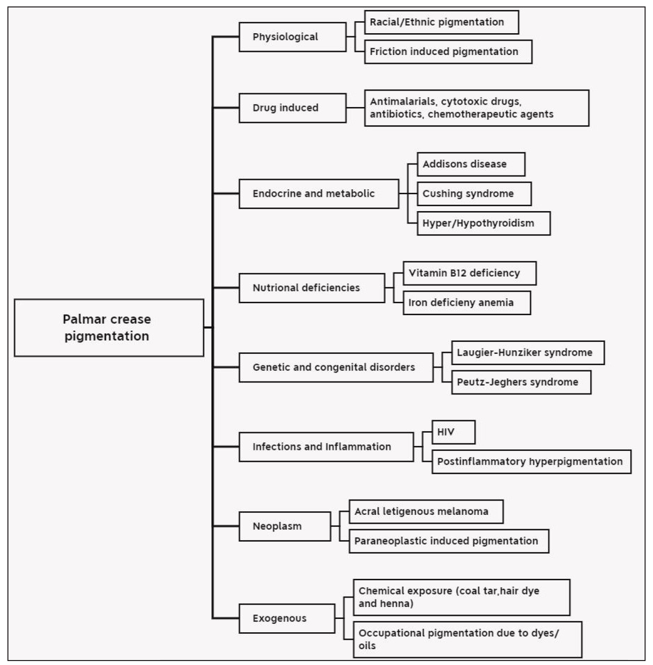
5. Palmar crease hyperpigmentationPalmar crease pigmentation [Figure 5] refers to darkening or discolouration along the palmar creases due to various physiological, pathological, or external factors. It may manifest in systemic diseases, nutritional deficiencies, or exposure to chemicals. Various causes of palmar crease pigmentation have been listed in Figure 6.14
Export to PPT
Export to PPT
6. Palmar pitsPalmar pits are small, asymmetrical depressions on the palms and soles due to stratum corneum loss. They appear flesh-toned to pink, measuring 2–3 mm in diameter and 1–3 mm in depth.15 Palmar pits are classically associated with disorders such as pitted keratolysis, arsenical keratosis, Darier disease, and acrokeratosis verruciformis of Hopf.16
7. Palmar xanthomaPalmar xanthomas, or xanthoma striatum palmare, are plane xanthomas affecting the palms and finger creases, presenting as yellow-orange subcutaneous lesions. It is associated with dysbetalipoproteinemia.These lesions arise from microtrauma-induced lipid accumulation, linking localised inflammation to systemic lipid metabolism abnormalities.17
8. Punctate porokeratosisPunctate porokeratosis (PP) is an uncommon variant of porokeratosis, first identified by Rahbari et al. in 1977. It presents as small, seed-like lesions with raised margins, appearing either in a linear distribution or diffusely across the palms and soles.18
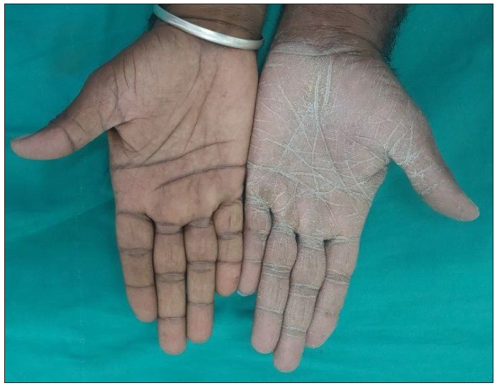
9. Tinea manuumTinea manuum is a dermatophyte infection of the palms, often presenting with diffuse scaling and hyperkeratosis [Figure 7]. Dermoscopy aids diagnosis by revealing whitish scales along palm creases, distinguishing it from other dermatoses.19
Export to PPT
10. Tinea nigraTinea nigra is a superficial fungal infection of the stratum corneum caused by the dematiaceous fungus Hortaea werneckii. Tinea nigra presents as dark brown or black patches on palms and soles, with accentuated pigmentation along palmar creases.20
Palmar crease pigmentation scaleThe palmar crease pigmentation scale predicts the risk of post-inflammatory hyperpigmentation (PIH), a genetically determined reactive melanosis independent of skin phototype or eye colour. The 4-point visual scale evaluates pigmentation contrast between palmar creases and surrounding skin: score 0 (no difference), Score 1 (low difference), score 2 (moderate difference) and score 3 (high difference). Scores 0 and 1 indicate low PIH risk, while scores 2 and 3 suggest higher risk.21
Comments (0)