RA is a lifelong condition associated with serious complications including progressive disability, and premature death. Its effects extend beyond the joints to include other body parts such as skin, eyes, lungs, and heart (Gad et al. 2021; Jang et al. 2022b). Current anti-rheumatic agents are only capable of managing symptoms and slowing down disease progression (Allen et al. 2018), which entails the continuous search for alternative treatments. DAPA has been reported in several studies to possess lots of pharmacological activities, including anti-inflammatory, apoptotic-modulation, and autophagic enhancement (Abdollahi et al. 2022; Anton et al. 2023; Arab et al. 2021; Deger et al. 2022; Jang et al. 2022a, b). In the present study, we introduced the possible therapeutic effects of DAPA against the AIA model in rats and the underlying mechanisms behind these outcomes, focusing on autophagy and Hedgehog pathways involvement.
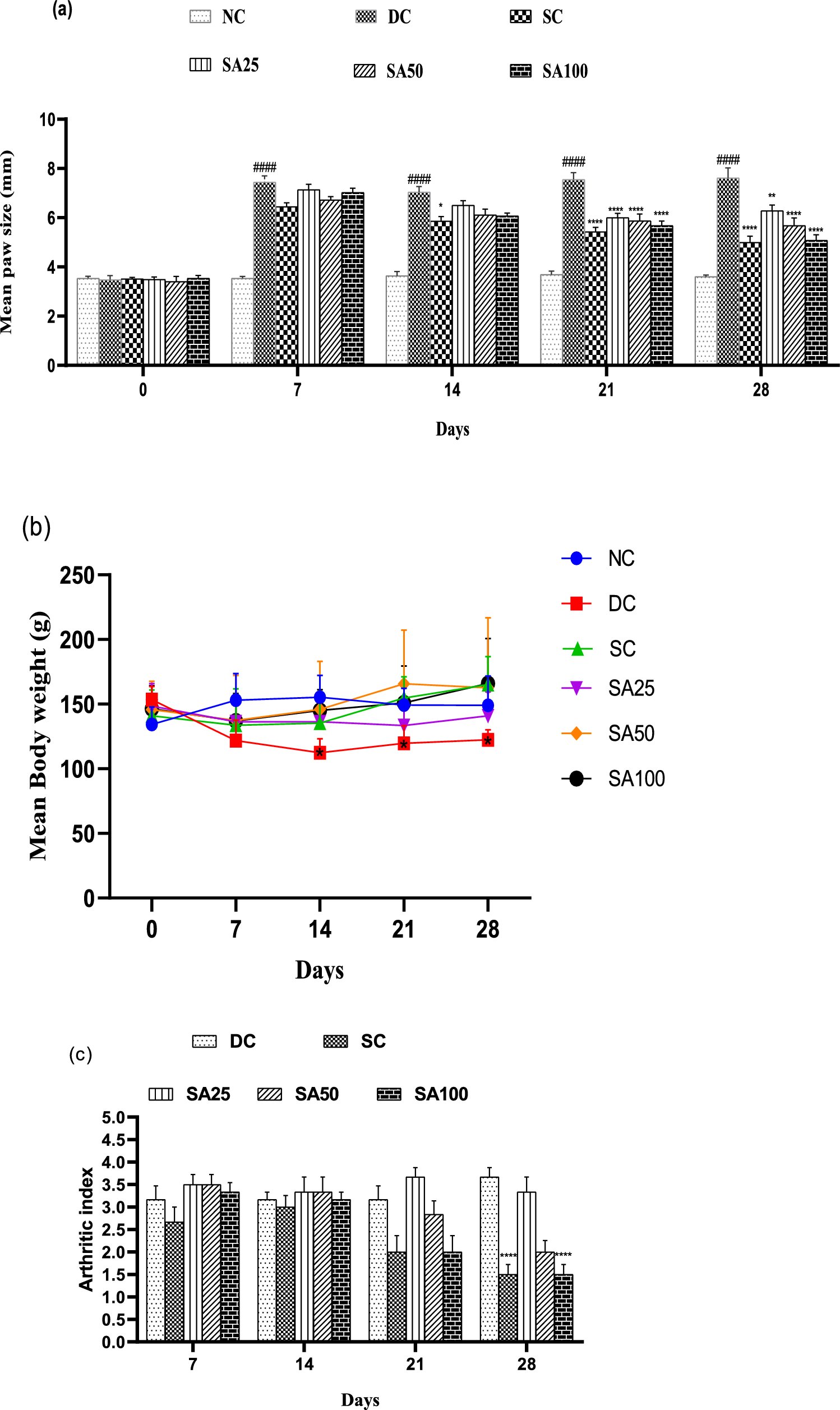
In the current work, an animal model of arthritis was well established after 24 h of injecting CFA, in which treated and non-treated rats exhibited significant inflammatory and arthritic alternation, which were boosted by the successive injections of CFA. This was reflected by the weakened locomotion of AIA rats as a result of their hind paws' severe edema and the apparent degrees of redness, swelling, and limited activity. The arthritis induction was supported by histopathological analysis of articular tissues obtained from the arthritic non-treated rats which demonstrated multiple focal articular surface irregularities, accompanied by diffuse inflammatory cell infiltrates in synovial membranes and periarticular connective tissue. These findings were consistent with earlier studies showing similar deteriorated arthritic parameters in CFA-induced arthritis models (Gaafar et al. 2018; Gad et al. 2021).
As part of our research, we conducted a preliminary dose–response study to determine the potential therapeutic benefits of DAPA against AIA-model in rats using three different doses (1, 5, or 10 mg/kg/day). The lower doses of DAPA (1 or 5 mg) revealed slight alleviation in arthritic parameters, while the highest dose (10 mg) presented eminent antiarthritic effects of reduced paw diameter, gait score, and AI, compared to lower doses. This novel discovered effect of DAPA halted the multiple histopathological abnormalities incited by CFA in a dose-dependent manner, preserving almost normal intact articular surface upon using the highest dose, without apparent abnormal alterations nor inflammatory cell infiltrates. These results of our study provide the first evidence for the promising antiarthritic impact of DAPA in an adjuvant-induced rheumatoid arthritis rat model. Based on these notable findings, we were encouraged to conduct further investigations to unmask the underlying antiarthritic mechanism of DAPA, in favor of the maximal used dose (10 mg/kg). Consequently, the second research phase was carried out to ascertain the efficacy of DAPA in treating RA, either alone or in conjunction with MTX as an established RA standard medication, along with exploring the possible involvement of the Hedgehog pathway as a valuable area of interest.
CFA has been extensively used to induce arthritis in experimental animal studies which mimics human rheumatoid arthritis. In the present study, administration of CFA-induced arthritic alternation confirmed by elevated AI, observed paw inflammation and edema, increased gait score, and paw diameter compared to healthy non-arthritic rats. These results agreed with previous studies reporting the arthritic effects of CFA in rats and mice (Zhu et al. 2020a; Weng et al. 2021; Tran et al. 2023). According to our results, monotherapy of either MTX or DAPA slightly reduced both AI and gait score of arthritic rats, while notable improvements were found in paw diameter. The combination of DAPA with MTX supplemented a further enhancement in all arthritic indices, in comparison to both MTX or DAPA alone.
Upon radiographical inspection, administration of CFA displayed arthritic alternation in rats that appeared as soft tissue swelling, bone erosion, and joint space narrowing, in agreement with previous studies reporting CFA-arthritic radiographic effects (Patel et al. 2021; Sharma et al. 2023). Radiographic examination of the treated groups with MTX or DAPA showed no discernible inhibition of the joint abnormalities associated with arthritis. Meanwhile, concomitant treatment of DAPA with MTX significantly improved arthritic changes in joint structure, particularly erosion, and joint space which was confirmed by histological examination.
In RA, MMPs including MMP-1, MMP-3, MMP-8, and MMP-13 were found to be a crucial element for joint health, disease development, and progression (Araki and Mimura 2017; Bian et al. 2023). Prior clinical investigations reported that patients with RA have elevated serum and synovial fluid levels of MMP-1 and MMP-3 compared to osteoarthritis patients and normal ones, correlated to the levels of other disease activity markers (Abdelrahman et al. 2020; Hussein & Aboukhamis 2023; Li et al. 2022; Yoshihara et al. 2000). According to research, MMP-1 and MMP-3 played key roles in the pathogenesis of RA and have been suggested as measures of disease progression and joint destruction (Wang and Khalil 2018). More specifically, in early-stage RA non-treated patients, MMP-3 levels have been associated with the prediction of disease progression and treatment outcome (Yeo et al. 2022; Hussein and Aboukhamis 2023). It was found that elevated MMPs and positive RF have been associated with higher bone tissue destruction, chronic inflammation, more nodules, and multisystem involvement (Lin et al. 2020; van Delft and Huizinga 2020; Petrovská et al. 2021). In accordance, our study revealed that compared to healthy non-arthritic rats, CFA significantly elevated serum levels of MMP-1, MMP-3, and RF. Interestingly, treatment with 10 mg of DAPA alone showed a dramatic decrease in these elevated serum levels in arthritic rats. Furthermore, these critical rheumatoid severity markers were further reduced by the combined regimen of DAPA with the conventional MTX medication compared to both arthritic non-treated rats and MTX treatment alone.
Several immunological and inflammatory mechanisms are activated throughout the development of RA, which is described as an immune-mediated inflammatory process. Joint degeneration in RA is caused mainly by an imbalance between the pro- and anti-inflammatory cytokines release (Guo et al. 2018; Ding et al. 2023). The current data showed that CFA multiple injections induced a systemic inflammatory state in diseased rats, illustrated by the heightened serum levels of NF-κB, and the subsequent inflammatory cascade of IL-1β, IL-6, and TNF-α, in alignment with recent works (Nazir et al. 2023; Dar et al. 2023). According to previous reports, oral administration of DAPA (5 and 10 mg/kg) has shown an anti-inflammatory activity via suppression of matrix metalloproteinases levels with significant inhibition in IL-1β, IL-18, and TNF-α levels in lipopolysaccharide-mediated lung injury (Abd El-Fattah et al. 2022). On the same track, we revealed that treatment with DAPA (10 mg/kg/day) alleviated the convinced systemic inflammation in arthritic rats, shown by the downregulated levels of NF-κB and the successive cascade of TNF-α, IL-1β, and IL-6. Surprisingly, the accretion of DAPA to MTX standard therapy in the current study showed an additional reduction in serum levels of all inflammatory cytokines. This finding supports the prevalent anti-inflammatory impact of DAPA, which would benefit RA treatment.
Previous studies concerning possible remission of rheumatoid arthritis showed that activation of AMPK plays a significant role in regulating inflammation and disease progression. Accordingly, AMPK activation in RA models showed enhanced inflammatory response via reduction of IL-6, modulation of immune response, and attenuation of cartilage damage which could be linked to subsequence inhibition of Hedgehog pathway, responsible for FLS migration and pannus formation (Guma et al. 2015; Li et al. 2015a, b; Wang et al. 2022). AMPK activation has also been linked to the downregulation of several inflammatory mediators including TNF-α, IL-1β, IL-6, and NF-κB along with the restriction of Gli-1 transcription factor activation in different animal models (Wu et al. 2019; Wang et al. 2022). Indeed, multiple models have linked DAPA to increased AMPK activity. According to Faridvand et al. (2022), DAPA pretreatment significantly reduced high glucose-induced apoptosis and CASP-3 activity in human umbilical vein endothelial cells through increased p-AMPK. In the same context, our results showed that CFA administration constrained the articular phosphorylation activity of AMPK, which explains the heightened inflammatory state in arthritic non-treated rats. Meanwhile, treatment with DAPA showed dramatically increased AMPK phosphorylation activity in the articular tissue of arthritic rats. Surprisingly, DAPA had added such a benefit to MTX treatment, in which the latter failed to significantly activate AMPK phosphorylation. These confirmatory findings might disclose the impediment effect of DAPA on NF-κB and the successive cascade of TNF-α, IL-1β and IL-6. Consequently, DAPA certified itself as a substantial anti-inflammatory agent, which might repurpose DAPA as a promising treatment for systemic inflammatory disorders such as RA.
Studies on RA have shown that one of the key pathophysiological factors, especially in cases of synovial hyperplasia and pannus development, is the reduced apoptotic sensitivity of synovial fibroblasts. (Ding et al. 2023). It was reported that in RA joint, FLS showed cancer-like behavior, characterized by aberrant proliferation and resistance to apoptosis, which in turn induced persistent inflammation and tissue damage (Mousavi et al. 2021; Wu et al. 2021; Xi et al. 2022). In the current study, administration of CFA significantly downregulated articular levels of CASP-3 and CASP-9 as well as articular gene expression of p53 and Bax/Bcl2 in arthritic non-treated rats compared to healthy non-arthritic ones. Interestingly, in comparison to MTX-treated rats, treatment with DAPA alone significantly elevated cartilage tissue levels of CASP-3 and articular gene expression of p53 while there was no recorded difference in CASP-9 and Bax/Bcl2 ratio. Our results also showed that administration of DAPA in conjugation with MTX restored impaired apoptosis through a significant increase of CASP-3 and CASP-9 articular levels as well as elevated p53 articular gene expression compared to MTX treatment alone. In contrast to untreated arthritic rats, the Bax/Bcl2 expression was substantially increased in the combined DAPA/MTX-treated rats. These findings agreed with earlier research, reporting DAPA therapeutic impact on a mouse solid tumor model via improving the survival rate, and CASP-3 activities (Kabel et al. 2021).
In RA, autophagy has been found to play a dual role both protective and pathogenic depending on the cell type and disease stage (Vyawahare et al. 2022; Liu et al. 2023). Previous studies reported that autophagy was involved in RA-FLS survival and resistance to apoptosis as well as T-cell activation, osteoclast differentiation, and bone resorption. On the contrary, autophagy was also linked to chondrocytes and cartilage homeostasis maintenance (Karami et al. 2020b; Guo et al. 2021). In the same context, recent studies have shown that induction of autophagy was associated with the suppression of arthritis severity and desirable therapeutic outcomes in vivo and in vitro (Fernández-Rodríguez et al. 2021; Wang et al. 2024; Yang et al. 2022).
According to Ibrahim et al. (2022a, b), DAPA demonstrated a prospective therapeutic benefit through modulating autophagy in an Alzheimer's rat model as evidenced by increased hippocampal gene expression of Beclin1. In our study, the administration of CFA dramatically lowered the articular gene expression of ULK-1, Beclin-1, and ATG-7 as compared to control rats, leading to significant downregulation in the autophagy process, supporting the hypothesis of autophagy involvement in hindering arthritis severity. With the same concept, our investigation revealed that treatment with DAPA activated the autophagy axis, as evidenced by notable elevation of articular autophagic markers ULK-1, Beclin-1, and ATG-7 gene expression. Moreover, the adjuvant therapeutic regimen of DAPA with MTX dramatically elevated articular gene expression of ULK-1, Beclin-1, and ATG-7 compared to either arthritic non-treated or MTX-treated groups. Collectively, in the current study, DAPA exerted a dual beneficial effect through enhancing autophagy as well as modulating aberrant apoptosis in the RA model in a similar behavior to its effect against tumor models. This precious impact of DAPA in restoring the haemostatic balance between apoptosis and autophagy pathways in RA would help to prevent aberrant and uncontrolled growth of diseased cells, which is the primary factor in the seriousness of arthritis, and degeneration of joints.
Hedgehog signaling pathway has been linked to RA pathogenesis, specifically through abnormal proliferation and invasion of FLS in the synovium (Su et al. 2022; Zhu et al. 2022b). Studies reported that aberrant activation of Hedgehog pathway in RA patients contributed to RA-FLS multiplication and migration in a tumor-like behavior leading to synovial inflammation, joint destruction, and pannus formation through different mechanisms including activation of MAPK/Jun N-terminal kinase pathway, MAPK/extracellular signal-regulated kinase pathway, and upregulation of MMP1 and MMP3 which plays a crucial role in FLS aggressive behavior (Zhu et al. 2020c; Mousavi et al. 2021). According to research, overexpressed Hedgehog signaling components including Ptch1, Smo, and Gli1 were found in RA synovial tissues and FLS cultures (Su et al. 2022; Wang et al. 2014). Following the same lines, our results demonstrated that arthritic non-treated rats significantly elevated Shh, Smo, ptch1, and Gli-1 articular gene expression. Fortunately, treatment with DAPA either alone or in conjugation with MTX dramatically suppressed articular gene overexpression of Shh, Smo, ptch1, and Gli-1 when compared to MTX alone. These results clarify that DAPA mitigates RA pathogenesis by inhibiting Hedgehog pathway overactivation, an important factor in the disease's progression, contributing to joint inflammation and cartilage destruction.
Targeting the crosstalk of signaling pathways in multiple disorders, notably RA, is a promising approach for the evolution of novel therapeutic agents. AMPK, the substantial regulator of the inflammatory cascade, plays a core role in the crosstalk between autophagy and apoptosis in different pathological states (Villanueva-Paz et al. 2016). AMPK is conceivably linked with different autophagy stages. On the other hand, it has been suggested that AMPK regulates cell apoptosis under stressful circumstances. The phosphorylation of AMPK subsequently leads to the stimulation of proapoptotic mediators p53 and Bax, thus encouraging cells to go through apoptosis (Villanueva-Paz et al. 2016). Furthermore, AMPK is also linked to the regulation of Hedgehog signal in previous studies. One of the SGLT2 inhibitors induced AMPK phosphorylation, which hindered the Hedgehog pathway expression, thus restoring apoptosis and preventing abnormal cancer cell proliferation (Xie et al. 2020).
By highlighting our findings, DAPA has strongly targeted AMPK, the key controller of the inflammatory cascade. DAPA halted the CFA-induced inflammatory state of NF-κB, and the downstream cascade of TNF-α, IL-1β, and IL-6. According to recent studies, DAPA has been reported to suppress inflammation and enhance autophagy through activation of AMPK phosphorylation in different animal models (Hassan et al. 2024; Ibrahim et al. 2022b; Luo et al. 2023). Herein, DAPA activated AMPK-phosphorylation with subsequent enhancement of the autophagy process, via elevated articular expressions of ULK-1, ATG-7, and Beclin-1. Our study revealed that DAPA-induced AMPK effect has led to restoring apoptosis in arthritic tissue, through CASP-3, CASP-9, and p53 articular activation, as well as boosting Bax/Bcl2 ratio. Additionally, DAPA-mediated phosphorylation of AMPK might engage in its inhibitory action of Hedgehog signal, illustrated by Gli-1, Smo, and ptch1 articular downregulation. All of these beneficial impacts of DAPA were reflected in the improvement of AI, gait score, and paw swelling, along with the reduced levels of RF, MMP-1, and MMP-3 arthritis markers. Therefore, DAPA improved the histopathological deteriorations in articular tissues exerted by CFA administration.
During the current study, we aimed to clarify the possible antiarthritic effects of DAPA monotherapy, in addition to the potential beneficial impact of combining DAPA with MTX as an established drug for RA treatment. MTX has been considered the most conventional therapy for RA since the 1980s till this day and is often called the first-line medication for RA treatment, regardless of the serious limitations associated with long-term use, such as liver damage, infection, and developing lymphoma. Therefore, MTX was used in this study as a standard therapeutic reference for the treatment of RA. The mechanism underlying MTX effectiveness in RA was previously described by several theories. These include antagonistic effects of folate, adenosine signaling, oxidative stress induction, reduction of adhesion molecules, modification of cytokine profiles, and suppression of inflammation. The anti-inflammatory outcome of MTX only referred to the upregulated adenosine levels (Friedman and Cronstein 2019). However, the impact of MTX on serious signaling pathways involved in RA, such as Hedgehog and autophagy, and the exact underlying anti-inflammatory molecular mechanism, was not clearly defined.
Previous studies concerning MTX reported significantly lowered blood levels of MMP-3 with no evidence of MMP-1 levels in RA patients (Green et al. 2003; Yeo et al. 2022). Upon comparing the antiarthritic mechanism between MTX and DAPA monotherapies, we found that DAPA significantly improved serum measures of MMPs and RF, leading to normalizing these markers in combination therapy. Curiously, DAPA monotherapy prevailed in superior actions in impeding inflammatory cascade in comparison to MTX monotherapy towards all the inflammatory mediators assessed in the current work, specifically through increased AMPK phosphorylation activity in the articular tissue of arthritic rats, adding such a benefit to MTX treatment, in which the latter failed to achieve it. Such advantage made MTX benefit from a combination regimen with DAPA, giving the combination therapy the superior anti-inflammatory impact over both monotherapies.
While most MTX studies in RA revealed activated JNK pathway and subsequent induction of p53 gene expressions, there are no precisely detailed studies associated with its effect over other apoptotic markers (Spurlock et al. 2011, 2012; Wang et al. 2020a). During the current study, DAPA achieved more prominent autophagic and apoptotic effects than MTX in CASP-3 and P53 articular expressions and all autophagic assessed parameters. Our analysis revealed that combining DAPA with MTX has earned MTX further autophagic and apoptotic activity against invasive RA tissues, targeting the two crossed pathways responsible for cartilage and joint degradation. The current result is considered a new finding for MTX autophagic activity, which came in contrast with a previous study that showed reduced mRNA and protein levels of autophagy-related genes of ATG (3, 5, 12), ULK1 and Beclin1 in MTX-treated arthritic rats (Sun et al. 2023).
A previous study reported the possible impact of MTX on the Hedgehog pathway when only combined with curcumin for osteosarcoma treatment, not for RA (Giliberti et al. 2024). Herein, DAPA monotherapy achieved dominant enhancement in this serious pathway in correlation with MTX alone. Additionally, combining DAPA with MTX gained both monotherapies further improvement in Hedgehog signal, supporting the previous study finding and offering a valuable therapeutic benefit via modulating the pathway responsible for the abnormal multiplication of FLS in RA.
In conclusion, our research revealed the first evidence for the ameliorative effects of DAPA against CFA-induced arthritic model in rats alone or adjuvant to conventional RA therapy. DAPA has certified itself as a multitarget agent by aiming at AMPK, the hallmark of the interplay between apoptosis and autophagy, as well as Hedgehog pathways. These newly discovered antiarthritic impacts of DAPA could be attributed to its anti-inflammatory, autophagic, and apoptotic modulation properties, interlined with curbing the Hedgehog signaling parameters. Nevertheless, we recommend further investigations on the possible corroboration of DAPA in RA patients and other related inflammatory disorders, as monotherapy or as adjuvant to the conventional regimens.


Comments (0)