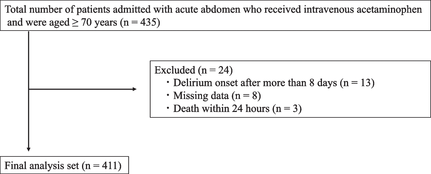
Hypocalcemia is one of the most frequent electrolyte disorders associated with anti-EGFR monoclonal antibody therapy. According to a pooled analysis, all-grade hypocalcemia caused by cetuximab was observed in 16.8% of cases, and grade 3/4 symptoms induced by either cetuximab or panitumumab were reported in 3.8% of all cases [10]. The general mechanisms of anti-EGFR treatment-induced hypocalcemia are multifactorial and associated with hypomagnesemia [9]. Impaired magnesium-dependent adenyl cyclase generation by cAMP reduces the release of parathyroid hormone, and skeletal resistance to this hormone during magnesium deficiency has also been implicated [9]. Additionally, hypomagnesemia alters the normal heteroionic exchange of calcium and magnesium at the bone surface, leading to increased bone release of magnesium ions in exchange for increased skeletal uptake of calcium from the serum [9]. Thus, hypocalcemia during anti-EGFR therapy is considered secondary to hypomagnesemia. Maliakal et al. suggested that hypocalcemia is a classic sign of severe hypomagnesemia (< 1.2 mg/dL) [9]. However, in the current case, we observed independent hypocalcemia without hypomagnesemia or notable serum magnesium variation, considering the electrolyte changes during each treatment cycle; however, several laboratory data were missing. The concomitant medications described in Table 1, except lansoprazole, were not associated with hypocalcemia, and lansoprazole was administered during the third cycle alone. Thus, we considered the symptoms observed in the current case were due to cetuximab sarotalocan and assessed the relationship using the Naranjo Adverse Drug Reaction Probability Scale, with a score of 6, indicating a"Probable"association (Supplemental Table 1).
Although there have been previous reports of electrolyte abnormalities associated with cetuximab sarotalocan, hypocalcemia has not been reported [11]. Furthermore, it remains unclear whether the reported electrolyte abnormalities are attributable to cetuximab or sarotalocan components. Although hypocalcemia has been reported with cetuximab alone, no such reports exist for cetuximab sarotalocan; therefore, we hypothesized that cetuximab caused the observed hypocalcemia.
Interestingly, the reduced serum potassium levels occurred in parallel with serum calcium levels, whereas serum sodium and chloride levels did not. In addition, a decrease in serum potassium levels can be occasionally observed in patients administered anti-EGFR monoclonal antibodies, typically secondary to hypomagnesemia [9]. However, the findings in our case differed, suggesting the occurrence of independent hypocalcemia and hypokalemia apart from hypomagnesemia.
Thomas et al. have reported a similar case of independent hypocalcemia associated with cetuximab; a patient post parathyroidectomy without renal impairment received cetuximab and irinotecan for metastatic colorectal cancer therapy and exhibited severe hypocalcemia after one week of chemotherapy (decrease from 7.0 to 5.7 mg/dL) [12]. She re-developed symptoms despite receiving oral calcium carbonate 1,000 mg TID and magnesium oxide 420 mg BID. Impressively, a 24-h urine evaluation revealed low calcium excretion and elevated fractional excretion of magnesium, although the serum magnesium levels were within the normal range [12]. Moreover, Chen et al. reported two cases of cetuximab-induced refractory hypokalemia without hypomagnesemia [13]. A patient who underwent a urine examination exhibited a non-obvious increase in potassium excretion, similar to the hypocalcemia reported previously [12]. Based on these facts, the temporary decrease in serum calcium levels in the current case may be due to reduced calcium absorption and/or abnormalities in bone metabolism and not due to enhanced excretion; however, renal calcium excretion was not assessed in our patient.
This patient had developed hypoparathyroidism upon undergoing subtotal thyroidectomy, and substantially reduced serum calcium levels and hypocalcemia were observed after the first administration. The dosage of alfacalcidol was reduced on days 133 and 140, and serum calcium levels decreased after dose reduction, regardless of cetuximab sarotalocan administration. Additionally, calcium intake, particularly on day 2, was lower than usual in the first cycle, during which a notable decrease in serum calcium levels occurred. These results suggest that calcium intake possibly impacted serum calcium reduction after administration in the current case; however, it remains unclear whether reduced intake due to invasive tumor illumination or uptake inhibition by cetuximab sarotalocan influenced these results. Moreover, the lower serum calcium variations observed in the second and third cycles could be attributed to the lower impact of calcium intake owing to reduced alfacalcidol dosage, resulting in its reduced uptake.
Hypocalcemia is a typical symptom of hypoparathyroidism [14]. Serum calcium levels decrease 4 h after thyroidectomy, suggesting that calcium levels decrease rapidly without supplementation [15]. A previous case report documented the same symptoms with cetuximab treatment, with the patient having previously undergone parathyroidectomy [12]. Consequently, concomitant hypoparathyroidism, in addition to reduced calcium intake, may have contributed to the observed decrease in this patient (Fig. 1, Fig. 2), as it is unlikely that a substantial decrease in serum calcium levels occurred due to reduced calcium intake for a few days (Fig. 3) alone, from a homeostasis perspective. Furthermore, the patient exhibited moderate renal impairment, which may have affected the results, although the renal involvement may have been low.
When administering anti-EGFR monoclonal antibodies to patients with hypoparathyroidism, pharmacists should proactively request serum electrolyte tests, including calcium levels, to enable the early detection of hypocalcemia. It is also important to educate patients about the possible symptoms and potential delays in calcium recovery. Additionally, assessing total calcium intake from medications, enteral nutrition, and diet is a key aspect of pharmaceutical care.
In general, intravenous or oral calcium supplementation and adjustment of serum magnesium levels are performed to treat anti-EGFR monoclonal antibody-induced hypocalcemia [9, 16]. Active vitamin D supplementation is pivotal for managing chronic symptoms in patients with hypoparathyroidism [16]. In the current case, grade 2 hypocalcemia disappeared without medication, possibly due to a single administration of the suspected agent during a certain period. Conversely, the possibility of rebound hypercalcemia after several weeks of hypocalcemia treatment has been reported [17], as observed in the current case, implying the importance of regular monitoring for early detection and intervention during the treatment, regardless of drug holidays. It is crucial to successfully manage the symptoms using the aforementioned methods in patients receiving cetuximab sarotalocan, as well as treatments with other anti-EGFR monoclonal antibodies.
This case report has several limitations owing to clinical practice. First, several data values were unavailable. The variation in serum electrolytes in the case of data missing, given the variation in the other treatment cycles, may have resulted in a discrepancy from the true results. Second, the renal excretion of electrolytes was not assessed. Third, genetic backgrounds were not evaluated. Consequently, our findings should be interpreted with consideration of these limitations.
In conclusion, we report temporary independent hypocalcemia without hypomagnesemia in a patient with hypoparathyroidism who underwent NIR-PIT using cetuximab sarotalocan. Regular evaluation of serum electrolyte levels is crucial for early and appropriate symptom management in patients receiving this treatment.


Comments (0)