Myopia is the most common refractive error globally, with an estimated half of the world’s population expected to be affected by 2050, and nearly 10% anticipated to have high myopia [1]. The prevalence of myopia and high myopia has shown a rapid increase, particularly in young East and Southeast Asians [1, 2]. The Implantable Collamer Lens (ICL; STAAR Surgical, Nidau, Switzerland) with a central hole facilitates the surgical procedure and safely and effectively corrects high myopia, super high myopia, astigmatism, and presbyopia [3,4,5,6].
Proper ICL sizing with an appropriate postoperative vault is vital for the success of the procedure. The consensus is that the optimal vault, which is the Distance between the posterior surface of the ICL and the anterior surface of the crystalline lens, should range from 250 µm to 750 µm7. Despite the manufacturer's provision of an online calculation for ICL sizing based on the white-to-white (horizontal corneal diameter, WTW) and anterior chamber depth (ACD), a significant proportion of cases still fall outside the optimal vault range [7, 8]. Consequently, many formulas and preoperative anatomic parameters have been proposed to optimise the method of ICL sizing [9,10,11]. Given the complexity of anterior segment biometrics, predicting the vault and ICL size based on preoperative parameters identified in previous studies remains problematic.
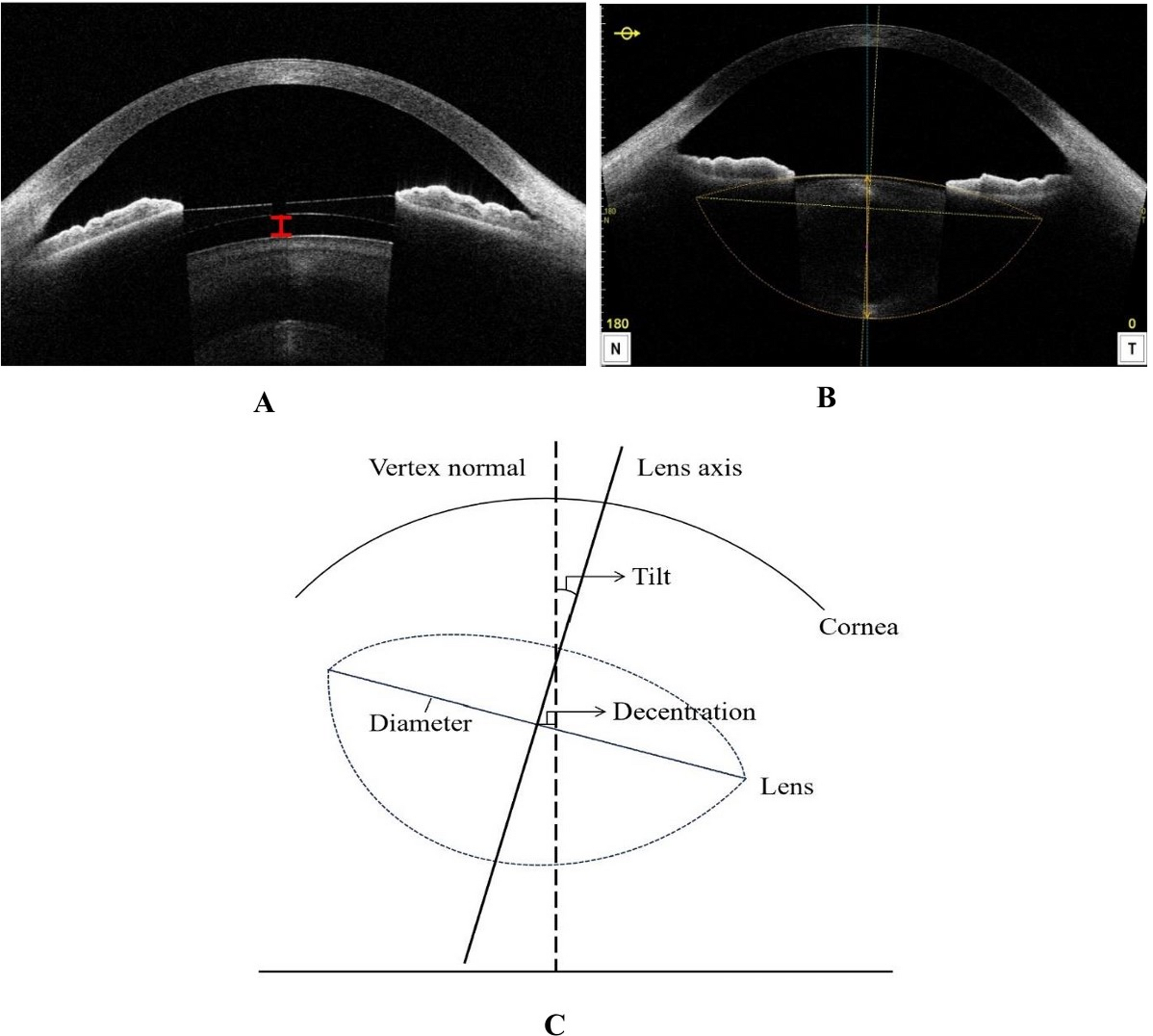
Theoretically, the ICL is positioned anteriorly to the crystalline lens and settles in the ciliary sulcus. However, current study has observed that the shape of the crystalline lens in high myopia tends to vary [12]. To improve the accuracy and precision of vault prediction, variables of the crystalline lens (e.g., crystalline lens rise, lens thickness, lens position, and lens surface curvature radius) have been introduced and are considered to be correlated with the vault [13,14,15]. The measurement of lens parameters depends directly on images scanned by high-frequency ultrasound biomicroscopy (UBM) and anterior segment optical coherence tomography (AS-OCT). However, UBM is invasive and shows poor reproducibility due to unstable measurement sites. In contrast, AS-OCT is widely used to image the lens and anterior part of the eye, owing to its non-invasiveness and enhanced accuracy. Recently, a new generation of swept-source AS-OCT (CASIA 2, Tomey Corporation, Nagoya, Japan) has been used to assess detailed crystalline lens characteristics in a three-dimensional manner [16]. This method can calculate detailed dimensional parameters related to the crystalline lens, including the anterior and posterior curvatures, the diameter, the decentration, and the tilt of the lens.
The present study intended to describe the lens morphologic features in Different vault at 3 months postoperatively using AS-OCT. Subsequently, we investigated the vault effects of the crystalline lens characteristics in highly myopic eyes following ICL V4c implantation.



Comments (0)