
Remember me


Hemorrhage control is a critical aspect of emergency medical care globally, as uncontrolled bleeding can contribute significantly to patient morbidity and mortality. Topical hemostatic agents are designed to rapidly control bleeding by promoting clot formation at the wound site. Various mechanisms are utilized by these agents, including mimicking the natural clotting process and creating physical barriers to reduce ongoing bleeding [6].
Fibrin-based sealants have gained popularity among modern hemostatic agents. One example is Evicel® (Ethicon Inc, Rariton, NJ), which emulates the natural blood-clotting process by providing fibrinogen and thrombin directly to the wound site. Fibrin and thrombin work together to form a strong, stable clot with adequate tensile strength to halt further bleeding [7]. Evicel® is unique in that it can be sprayed directly onto the wound, making for especially quick and easy application. The efficacy of Evicel® was recently studied among pediatric surgery patients to treat mild to moderate intraoperative bleeding. In this study, Evicel® required an average of four minutes to achieve hemostasis. This study compared treatment failure rates to Surgicel™ (Ethicon Inc, Rariton, NJ), an absorbable hemostatic agent, and found that treatment failure rates were 5% for Evicel® vs. 25% for Surgicel™. The study concluded that Evicel® can be safely used to control mild to moderate intraoperative bleeding [8].
In contrast to fibrin-based sealants, several chitosan-based sealants (i.e., HemCon® (Tricol Biomedical, Inc, Portland, OR), Axiostat® (Axio Biosolutions, India), and Celox® (Medtrade Products, Ltd. Crewe, UK)) can be used to treat acute bleeding. Chitosan is a sugar derived from the outer skeleton of shellfish (i.e., crab, lobster, shrimp), which is often combined with a synthetic or natural polymer to enhance its water resistance and thermal stability. Although chitosan is derived from shellfish, production of these sealants removes the tropomyosin allergen deemed responsible for allergic reactions to shellfish, and at least one study has suggested that these products can be safely used on patients with a shellfish allergy [9].
HemCon®, a commonly used topical hemostatic agent works by enhancing aggregation of red blood cells, stimulating platelets and altering the structure of fibrinogen [10]. In contrast, Axiostat® is a hemostatic dressing that uses the positively charged chitosan to attract negatively-charged platelets to form a clot. Celox® works similarly to HemCon® and Axiostat® as it is a dressing that contains chitosan-based granules that can be applied to a wound. One recent study compared the efficacy of Celox®, Algan hemostatic agent (AHA) powder, and control (i.e., saline soaked gauze) for bleeding cessation following femoral incision in rats. This study found that saline-soaked gauze was unsuccessful in stopping bleeding in all seven subjects. In contrast, Celox® was able to control bleeding and achieve hemostasis for all seven subjects in less than two minutes [11]. Another study assessed the efficacy of Axiostat® in assisting in the compression closure of femoral artery access sites following endovascular surgery. This study found that adequate hemostasis was able to be achieved in 91.7% of patients [12]. One recent meta-analysis found a statistically significant decrease in bleeding time when comparing hemostatic dressings including HemCon® versus control among anticoagulated patients undergoing dental procedures [13].
Xstat® (RevMedx, Wilsonville, OR) is another hemostatic agent frequently used for hemorrhage control. This agent is composed of cellulose sponges coated with chitosan, which quickly absorbs blood and expands volumetrically to provide internal wound compression. Xstat® is a unique agent because it is injectable, making it especially useful for the treatment of deep, penetrating wounds such as gunshot wounds [14, 15]. One recent study of swine subjected to liver hemorrhage (i.e., non-compressible torso hemorrhage) found that survival rates were significantly higher in the Xstat® group compared to controls [16].
Mechanical Closure DevicesModern mechanical closure devices use novel designs to circumvent some of the challenges traditionally associated with other closure methods, including prolonged application times, scarring, and pain with movement during healing. They also aim to minimize edge trauma, which is damage caused to the perimeter of the wound by excessive tension from sutures or staples, improper application technique, or simply by skin penetration. Some of these novel devices are designed to completely avoid skin penetration, thus minimizing this common cause of wound edge trauma and improving final scar cosmesis. These devices are especially useful for low-tension linear wounds, and offer quicker application than traditional methods, often without anesthesia. Two such devices, Zip® Surgical Skin Closure (Stryker Corp., Kalamazoo, MI) and DermaClip® (DermaClip LLC, El Paso, TX), both adhere to intact skin adjacent to the wound with an adhesive strip, utilizing a strap bridge along the length of the wound to apply tension and approximate the wound edges [17, 18].
DermaClip® (Fig. 1) is one of the most widely-used needle-free closure devices. Its strap bridge is composed of polypropylene tabs on either side which lock onto each other, lifting the wound edges and creating an everted wound edge, thus relieving wound tension and avoiding the negative sequelae associated with an inverted scar [18]. DermaClip® has been found to be especially useful in elderly patients, who frequently present with traumatic skin and tissue injuries and whose fragile, thin skin presents a unique challenge for traditional suture or staple application [18]. DermaClip® also requires less skill than traditional sutures and is inexpensive costing approximately $3.67 per centimeter [18]. The Zip® Surgical Skin Closure system (Fig. 2) has also been shown to be associated with less pain during movement following total knee arthroplasty (TKA) when compared to staples, likely due to more uniform distribution of mechanical support across the wound [17]. This system may also provide better scar cosmesis than staples when used for skin closure [19].
Fig. 1
DermaClip® (DermaClip LLC, El Paso, TX) application. Image used with permission from the manufacturer [20]. All rights reserved, DermaClip LLC 2025
Clozex® (Clozex Medical, West Bridgewater, MA) (Fig. 2) consists of multiple adhesive interlaced filaments anchored by pulling tabs that applies tension for wound closure. In one study, closure with Clozex® was found to be significantly faster to achieve than a running 3 − 0 Prolene suture post total knee arthroplasty (TKA) (266 vs. 91 s; P =.005) [21]. In this study, the Stony Brook Scar Evaluation Scale (SBSES) was also used to assess the appearance of wounds post-elective orthopedic procedures. The SBSES scores were shown to be significantly higher for wounds treated with Clozex® at 2 weeks when compared to suture (4.09 vs. 2.8; P =.005), indicating better overall scar appearance. This improved cosmesis was noted to persist at 3 months post-repair (3.82 vs. 2.85; P =.023) [21]. In addition, the cost of Clozex®, including unit cost and calculated operation room time, was significantly less than suture repair ($235.40 vs. $600.88 for an average wound size of 8.65 cm) [21].
Fig. 2
Clozex® (Clozex Medical, West Bridgewater, MA). Image used with permission from the manufacturer [22]. All rights reserved, Clozex Medical 2025
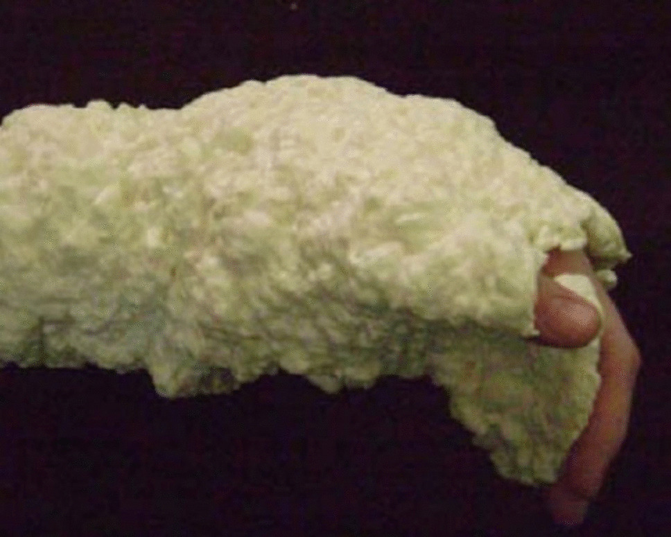
MicroMend® (KitoTech Medical, Seattle, WA) (Fig. 3) is another mechanical closure device that is easy to apply, resembling a butterfly bandage, and uses microstaples on either side of the lesion to provide tensile strength while minimizing edge trauma. It is available in various sizes and can close wounds ranging from 1-cm to 50-cm in length. It has been shown to produce significantly better aesthetic results than the use of traditional sutures. In one study evaluating portal closure methods, providers and patients rated wound appearance to be better with MicroMend® than with sutures in over 90% of cases and showed the device to be applied more easily and rapidly than sutures in 83% of cases [23].
Fig. 3
MicroMend® (KitoTech Medical, Seattle, WA). Image courtesy of the authors [24]
Negative Pressure Wound Therapy (NPWT)Negative pressure wound therapy (NPWT), also known as vacuum-assisted closure (VAC), utilizes a specialized wound dressing system to apply uniform, controlled sub-atmospheric pressure over a wound. These devices are most commonly used with complex and chronic wounds [25], although their utility has also been demonstrated for acute injuries. In the acute care setting, VAC may also be used for the stabilization of complex injuries, to facilitate safe interfacility transfer [26, 27]. This technology itself helps to facilitate wound healing by rapidly removing wound exudate, reducing the risk of bacterial colonization, promoting granulation tissue formation, and increasing peripheral microcirculation and oxygenation. In one study, 90% of NPWT-treated pressure ulcers healed after 43 days of treatment versus 48% of ulcers treated with conventional methods [28]. The use of NPWT has also been shown to improve closure rates for diabetic foot ulcers. In one study, 43.2% of patients achieved closure with a VAC system versus 28.9% of ulcers treated with advanced moist wound therapy [28]. The efficacy of NPWT has also been demonstrated in primarily closed surgical wounds. In one retrospective chart review, the use of NPWT was associated with a 22% rate of complications compared to 65.9% with traditional gauze dressings following abdominal wall reconstruction. In the same study, a lower rate of skin dehiscence occurred (9% vs. 39%) compared to dry gauze dressings [28]. Finally, VAC has been shown to increase the success rates of skin grafts. In one study, only 3% of VAC-treated wounds required a repeated skin graft, compared to 19% of those treated with bolster dressings [28].
Since the development of NPWT, a number of modalities have been developed to further increase efficacy, speed wound healing and minimize infection risk. One such modality is NPWT with instillation (NPWTi), consisting of the traditional VAC system with the addition of intermittent application of topical solution (i.e. regular saline, antiseptic solution) [29]. Compared to standard NPWT, NPWTi has been theorized to disrupt the biofilm via intermittent cleansing, dilute wound debris, and better promote the formation of granulation tissue, ultimately reducing bacterial burden and leading to greater reductions in wound size than NPWT alone [30]. In one study on patients with extremity ulcers, significant wound healing benefits of NPWTi over standard NPWT were demonstrated, including a mean reduction in wound size of 28.82 for NPWTi vs. 19.8 for standard NPWT (p <.05) [30]. In the same study, wound surface epithelium was evaluated and placed into three groups: improvement, mild improvement or moderate improvement. In the NPWTi group, there was found to be no-improvement in 1 (4.2%) out of 24 patients, mild in 13 (54.2%) and moderate in 10 (41.7%) compared to 7 (30.4%), 16 (69.6%) and 0 in the standard NPWT group (p <.001) [30]. The addition of instillation with dwell time to NPWT (NPWTi-d) has also been shown to improve wound bed cleaning and wound granulation [29]. In one study which incorporated a variety of surgical, chronic, and traumatic wound types, microbiological analysis of tissue samples taken before and after NPWTi-d with saline instillation showed a statistically significant reduction in the number of different bacteria from 2.38 to 1.16 (P <.05). A statistically significant reduction in bacterial count from 3.9 to 1.3 following NPWTi-d treatment (P <.05) was also demonstrated [29].
Advanced Adhesives and Tissue Bonding AgentsTissue adhesives include topical liquid sealants that can be used to close skin lacerations. They have gained popularity due to their quick and easy application, avoiding the need for skilled suturing techniques and eliminating the need for subsequent suture removal [31]. They appear to decrease both infection rates and wound closure times when compared to suture repair [32]. Cyanoacrylates are among the most widely used tissue adhesive groups and have evolved into modern formulations with improved properties. Newer generations of cyanoacrylates feature longer chemical chains, that further enhance their durability and reduce inflammatory effects on tissues. Both short- and long-chain cyanoacrylates are utilized in clinical practice, with their selection tailored to the type of wound and specific circumstances [33].
In the modern day, 2-octyl cyanoacrylate (i.e., Dermabond®, Ethicon Inc., Somerville, NJ), is used to close a wide array of different skin wounds [34]. In one study comparing wound healing using Dermabond® versus subcuticular suture closure following laparoscopic cholecystectomy, the authors found significantly greater wound closure speed in the Dermabond® group (i.e., 229.16 +/- 43.7 s vs. 258.82 +/- 51.7 s, p =.01). In the same study, wound breakdown rates and patient satisfaction scores were found to be similar [35]. A randomized controlled trial comparing wound healing in Dermabond® vs. subcuticular sutures following urogynecological procedures found that closure time was significantly decreased in the cyanoacrylate group (5.4 +/- 2.0 min vs. 24.9 +/- 5.6 min; p <.0005). This study also found no significant difference in wound cosmesis between the two groups [36].
Recent advancements in the field of tissue adhesives also include several new materials that may offer unique benefits. Light-activated adhesives are a type of tissue adhesive that uses light to activate its polymer material, allowing it to adhere to biological tissue. ChemPatch® (ChemMasters, Madison, OH) is composed of a fibrous material that reacts to near infrared radiation, promoting tissue repair and inhibiting proliferation of infection [37]. Other light-activated adhesives include self-luminescent porous silicon particles [38]. Of course, the effectiveness of those photosensitive tissue agents activated by direct sunlight is partially dependent upon the intensity of sunlight exposure [39].
HydrogelsHydrogels are gel-like substances consisting of polymer chains with the ability to absorb large quantities of water. As a result, they are porous, flexible and have a delicate structure that allows hydrogels to mimic the structure of living tissue [40]. Hydrogels are especially useful for expediting wound healing as they can absorb exudate, conform to irregularly-shaped wounds, provide antimicrobial and antibacterial properties, debride necrotic tissue, and provide a moist healing environment [41].
Hydrogels can also be used as add-ons to other wound closure methods. One advancing area of smart wound systems is drug delivery based upon biochemical sensors, including one method of delivering pharmaceutics through the use of “smart hydrogels” [42]. One such smart hydrogel (microgel@PAM/CS) is both temperature and pH-responsive and can serve as a drug delivery system for antibiotics paired with albumin. When compared to standard methods of drug administration (e.g., oral or intravenous injection), this hydrogel demonstrates much higher drug-loading efficacy [42]. At seven days post-treatment, subjects treated with the hydrogel appear to have reduced wound size without signs of inflammation, as well as overall improved wound healing when compared to controls [42].
Burn injuries are a common presentation to the ED and can have limb and life-threatening consequences. One common challenge with burns wound healing is the delivery of drugs across eschar to deeper tissues [43]. To combat this problem of drug penetration in burns, a smart chitosan-based hydrogel system has been developed. This system aids in the delivery of angiogenic material to the wound site with the aim of expediting wound healing. One study evaluating this system found that granulation tissue thickness was significantly greater in the group treated with hydrogel-based drug delivery systems when compared to the control group treated with a phosphate buffer solution [44].
Wound contracture is another area of special concern with burn-related wounds. One study examined rates of wound contracture in different hydrogel groups (i.e., gelatin-based hydrogel with cell spheroids, hydrogel only, hydrogel cell suspension only) versus a “no hydrogel” group. This study showed that the cell spheroid with hydrogel group had the highest rate of 14-day wound closure at 55.3%, followed by the hydrogel cell suspension group 45.2%, hydrogel only group at 37.1%, and no treatment group at 30.1% [45]. The authors concluded that hydrogels were not associated with any wound toxicity, and accelerated wound healing in rat models was supported by combining stem cells with hydrogels [45].
The efficacy of hydrogels has also been evaluated on a cellular level. One recent study found that wounds treated with a near-infrared (NIR) hydrogel developed more glands, fibroblasts, capillaries and collagen deposits when compared to the control group, as well as a tight affinity between the new dermis and epidermis, which was indicative of healing [46]. One amorphous hydrogel, EHO-85, has been shown to provide 26% higher average ulcer closure rates and three-times higher closure speed than controls at day 14 of treatment [47].
Biomarker Monitoring Wound Closure SystemsSome novel wound closure systems now allow for the monitoring of different biological parameters in order to ensure proper healing and rapid detection of biochemical irregularities [48]. These systems can transmit patient data to the healthcare provider for quick and easy access using electronic devices. Commonly monitored markers in wound care include physiologic parameters such as temperature, oxygen and moisture, or biomarkers such as pH, uric acid, and cytokines [49].
One example of a biomarker-monitoring system is a battery-less “smart bandage from Texas Instruments,” which is able to sense skin strain and temperature while being powered through the electromagnetic field generated from a near-field communication (NFC) electronic device. The information this “smart bandage” provides is able to be accessed by the user through a “SenseAble” software application [50]. Another available biosensor is the surface-enhanced Raman spectroscopy (SERS), a low-cost material device that is able to detect biologically relevant concentrations of TNF-alpha and matrix metalloproteinases. In one study involving a polypropylene mesh equipped with Raman spectroscopy, the device allowed for temperature measurement, which has proposed utility in the monitoring of post-surgical inflammatory states [51]. This type of smart wound dressing has the potential for use in the prevention of post-surgical bacterial infections.
Recent studies show the potential of smart wound dressings to inhibit fungal proliferation as well. One aggregation-induced emission luminogens (AIEgens)-based wound care system was developed with a fluorescent sensor and photosensitizer [52]. An image of this dressing taken on a smartphone could then be sent to a cloud software program to establish a diagnosis of Candida albicans infection beneath it. Photodynamic therapy (PDT) could also be delivered via the same smartphone if the necessary modifications to the device were in place. Although this proof-of-concept was completed in a mice model, diagnosis and treatment were achieved within minutes. Additionally, AIE-device-treated tissue recovered faster than with the commercial antifungal agent fluconazole, showing great potential in the treatment of fungal infections [52].
One of the key factors in creating a suitable environment for wound healing is avoidance of tissue hypoxia, which can otherwise provide a fertile environment for bacterial and fungal growth [53]. One study has actually demonstrated the ability of smart dressings to accurately detect tissue oxygenation levels using a flexible electrochemical biosensor [54]. Another factor in maintaining skin integrity is management of pH. The pH of healthy skin usually ranges from 4 to 6, but in dermal wounds the pH can become more alkaline, further increasing susceptibility to infection. Interestingly, smart wound systems may be able to detect and even treat pH abnormalities. One alginate-based dressing has been developed that is able to distribute antimicrobial particles when pH changes indicative of microorganism growth are detected [55].
Comments (0)