
Remember me

A total of 201 examinations from 169 patients less than 21 years of age were included. Patient mean age at examination was 12.1 (± 4.9) years. Ninety-two (54.4%) of the patients were female accounting for 106 (52.7%) of the examinations. Mean BMI was 21.6 (± 5.8) kg/m2 and mean BSA was 0.98 m2 with median BSA = 0.96 m2 (range 0.06–2.9). Twenty-four patients were six years of age or younger, all of whom had a BSA < 0.8 m2 and 59 other patients over the age of six had a BSA < 0.8m2. Fifty-five patients were sixteen years of age or older and 7 patients had a BSA > 2.2 (age range 12–20 years).
Fig. 1
Scatterplot of R1/R2 average measured secreted fluid volumes for patients by age of patient at examination. Diagnosis of patient is delineated. The dashed line indicates the 5th percentile threshold for secretion for age.
One hundred and thirty-six patients (67.7%) had clinical diagnoses of pancreatitis, 53 (26.4% of 201) of whom had chronic pancreatitis, 54 (26.9% of 201) of whom had acute recurrent pancreatitis, and 29 (14.4% of 201) of whom had acute pancreatitis. Frequencies of other diagnoses are detailed in Table 1. Average secreted volume between R1/R2 by age for patients is shown in Fig. 1. Twenty-nine patients underwent multiple examinations with secreted fluid volumes over time plotted in Fig. 2.
Fig. 2
Spaghetti plot of R1/R2 average measured secreted fluid volumes for patients that underwent multiple examinations. In most cases, secreted fluid volumes are relatively flat over time with a few patients showing pronounced increases or decreases
Table 1 Baseline characteristics of the study sample (n = 169). Values are means and standard deviations or counts and percentagesFrequencies of abnormal secretory responseTable 2 displays frequencies of abnormal secretory response based on automatically measured secreted fluid volumes and R1/R2 average secreted fluid volumes, subdivided by patient diagnostic group. Automatically measured secreted fluid volumes were abnormal based on BSA in almost twice as many cases (n = 135/201, 67.2%) as R1/R2 average volumes (n = 70, 34.8%). More patients had an abnormal secretory response based on BSA-derived thresholds than age-derived thresholds based on automatically measured secreted fluis volumes and R1/R2 average volumes (Table 2).
Table 2 Frequencies of abnormal secretory response by observer and judged by BSA or age-defined thresholds for various patient diagnostic subgroups. Results are counts (%) with 95% confidence intervals presented in brackets. R1 refined the automatically generated volumes and R2 further refined R1’s segmentationsAmong patients who had age or BSA outside of the range assessed by Trout et al., 20 of the 24 patients (83.3%) < 6 years of age had abnormal secreted fluid volumes based on the age-derived threshold for a 6 year old and 22/24 (91.7%) had abnormal secreted fluid volumes based on the BSA-derived threshold for a BSA of 0.8 m2 using R1/R2 average measured secreted fluid volume. Among the 55 patients who were ≥ 16 years of age, 6/55 (10.9%) had an abnormal measured secreted fluid volume based on the age-derived threshold and 8/55 (14.5%) had an abnormal measured secreted fluid volume based on the BSA-derived threshold.
Among the 81 patients who had a BSA < 0.8 m2 (ages 0–13 years), 50/81 (61.7%) had abnormal secreted fluid volumes based on the BSA-derived threshold while 54/81 (66.7%) had abnormal secreted fluid volumes based on age-derived thresholds. Finally, among the 7 patients who had a BSA > 2.2 m2, 2/7 (28.6%) had an abnormal measured secreted fluid volume based on the BSA-derived threshold and 0/7 (0%) had an abnormal measured secreted fluid volume based on the age-derived threshold.
Diagnosis-specific frequencies of abnormal secretory responseAmong the 136 patients with pancreatitis, R1/R2 average measured secreted fluid volumes resulted in characterization of secretory response as abnormal in 51 (37.5%) and 44 (32.4%) patients based on BSA and age respectively. Among subtypes of pancreatitis (acute, acute recurrent, chronic) frequencies were similar with overlapping 95% confidence intervals (Table 2).
Among the 17 patients with a clinical diagnosis of exocrine pancreatic insufficiency but without pancreatitis as documented in the patient’s chart by a pediatric gastroenterologist, 47% had an abnormal secretory response by BSA and age based on R1/R2 average volume.
R1/R2 average measured secretory response was classified as abnormal by both BSA and age in 29% (4/14) patients with clinical diagnoses of biliary disorders. Specific diagnoses associated with abnormal secretory responses included: bile salt export pump deficiency, two patients with anomalous pancreatobiliary junction, and biliary stricture.
For the patients with ‘Other’ clinical diagnoses, R1/R2 average measured secreted fluid volume was abnormal in one patient with pancreatic transection and one patient with history of pancreatic trauma.
Comparison to qualitative clinical assessmentClinical reports for the included MRI examinations characterized secretory response as qualitatively abnormal in 26/201 (12.9%) examinations. Compared to a reference standard of quantitative analysis based on R1/R2 average measured secreted fluid volume, qualitative assessment failed to identify an abnormal secretory response in 50 examinations based on BSA and 55 examinations based on age (false negatives) and falsely identified an abnormal secretory response in 12 examinations based on both BSA and age (false positives) (Table 3). Accuracy of qualitative assessment relative to the quantitative reference standard was 69% (95%CI: 62.3%−75.5%), sensitivity was 22% (95%CI: 12.5%−34.0%), and specificity 54% (95%CI: 36.4%−70.4%) using the BSA reference. Using the age reference, the accuracy of qualitative assessment was 67% (95%CI: 59.7%−73.1%), sensitivity was 20% (95%CI: 11.6%−31.7%), and specificity was 54% (95%CI: 36.4%−70.4%).
Table 3 Confusion matrix comparing qualitative characterization of secretory response via clinical reports for the MRI examinations as compared to a reference standard of quantitative analysis based on R1/R2 average measured secreted fluid volume. Accuracy of qualitative assessment relative to the quantitative BSA-referenced reference standard is 69% (95%CI: 62.3%-75.5%) with 22% (95%CI: 12.5%-34.0%) sensitivity, and 54% (95%CI: 36.4%-70.4%) specificity. Relative to the age-referenced reference standard, the accuracy of qualitative assessment was 67% (95%CI: 59.7%-73.1%) with 20% (95%CI: 11.6%-31.7%) sensitivity and 54% (95%CI: 36.4%-70.4%) specificitySecreted fluid volume measurement agreementThere was poor agreement between measured secreted fluid volume derived from the automatic segmentation and the segmentation refined by R1 (ICC = 0.38; 0.19–0.53 95% CI) with a mean difference in measured volumes of −29.0 mL (95% limits of agreement: −139.2 to 81.2 mL) (Fig. 3), reflecting an average underestimation of secretory response by the automated segmentation. There was excellent agreement between secreted fluid volume derived from the refined segmentation generated by R1 and the further refined segmentation by R2 (ICC = 0.97; 0.95–0.97 95% CI) with a mean difference in measured volumes of −1.7 mL (95% limits of agreement: −21.5 to 18.1) (Fig. 4). Overall, agreement between automatically measured secreted fluid volumes and R1/R2 average volumes was poor (ICC = 0.37; 0.17–0.52 95% CI) (Fig. 5) with a mean difference of − 30.1 mL (95% limits of agreement: (−141.6 to 81.4 mL).
Fig. 3
Bland-Altman difference plot for automatically measured secreted fluid volume versus R1-refined secreted fluid volume. Solid horizontal line indicates mean bias with dashed horizontal lines indicating the 95% limits of agreement. Short oblique dashed line reflects simple linear regression of the plotted data and suggests slight negative proportional bias reflecting decreased difference between the automatically measured volume and R1 refined measured volume as the secreted fluid volume increases
Fig. 4
A Scatterplot of secreted fluid volumes measured after R1 refinement of the automatic segmentation and R2 refinement of R1’s segmentation. The solid best-fit linear trend line and the dotted line of equality are nearly perfectly superimposed showing little disagreement between observers (ICC=0.96). B Bland Altman difference plot for fluid volume measurements based on refinement of the automatic segmentation by R1 and then subsequent refinement of the R1-refined segmentation by R2. Solid horizontal line indicates mean bias with dashed horizontal lines indicating the 95% limits of agreement. Short oblique dashed line reflects simple linear regression of the plotted data and suggests limited proportional bias
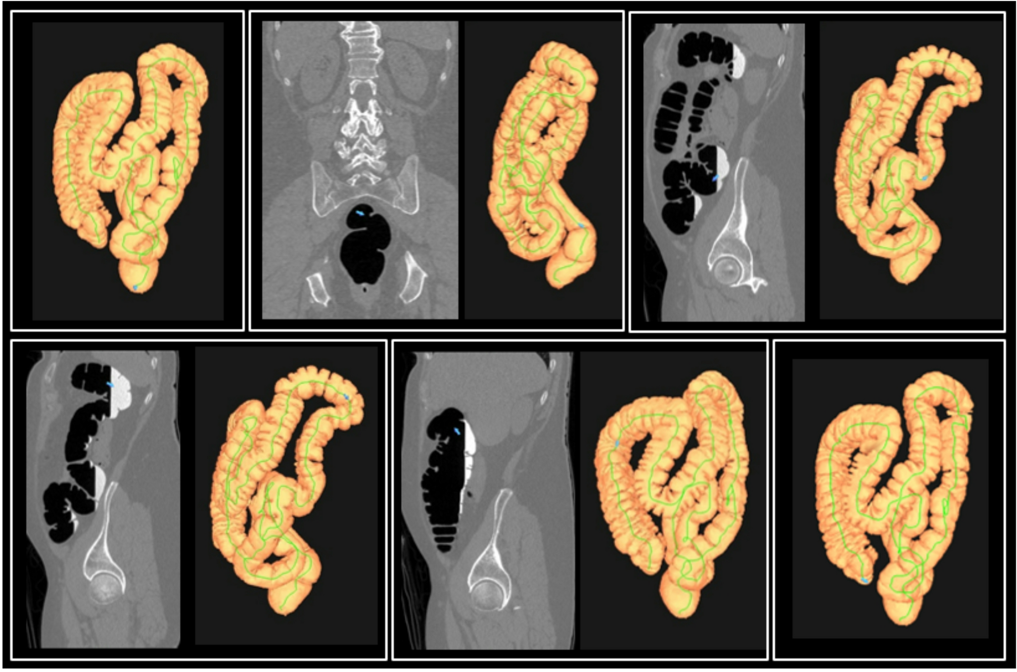
Processing timeProcessing time for R1 was captured for 191/201 data sets with the remainder not captured due to technical difficulties. The average time R1spent refining the automatic segmentation was 5.1 (± 3.2) minutes. Processing time for R2 was captured for 183/201 data sets and the average time spent refining segmentations already refined by R1 was 3.4 (± 1.8) minutes.
Fig. 5
Scatterplot of secreted fluid volumes measured using PFTquant (automatic segmentation) versus the average of volumes after R1 and R2 refinement. Each dot represents a different MRI examination. The solid line is the best-fit linear trend line and the dotted line is the line of equality. In general the automatically measured volume underestimates secreted fluid volume after human refinement (ICC=0.37).
Comments (0)