
Remember me

Approximately 4% to 30% of patients with POI have concurrent autoimmune disorders, with thyroid-related diseases, particularly autoimmune thyroid diseases (AITD), being the most strongly associated [4]. Therefore, early diagnosis and intervention of AITD are important for preserving ovarian function and improving reproductive health outcomes. Hashimoto’s thyroiditis, the most common form of AITD, has an estimated prevalence of about 18% in adult women [5]. AITD can also manifest as hyperthyroidism, notably in the form of Graves’ disease, which is characterized by abnormal thyroid hormone levels—either elevated or reduced—and the presence of thyroid autoantibodies [6]. Studies suggest that women with AITD are more likely to experience reduced ovarian reserve [7]. Thyroid peroxidase antibodies (TPO-Ab) are key biomarkers for diagnosing autoimmune thyroid diseases, and their presence is closely linked to impaired ovarian function [8]. Furthermore, histological examination of ovaries in POI patients often shows either complete follicular depletion or varying numbers of residual follicles. The latter is observed in approximately 40% of POI cases, which aligns with ultrasound findings that 41% to 60% of POI patients retain some residual follicles [9]. These findings emphasize the complex relationship between AITD and ovarian dysfunction, highlighting the need for further research into the role of ultrasound in evaluating ovarian health. A better understanding of this connection could significantly improve care and quality of life for women affected by AITD-related ovarian dysfunction.
Studies suggest that ovarian morphology in women with thyroid autoimmunity may show abnormalities on ultrasound [10]. For example, some women exhibit irregular follicular development, while others may show reduced ovarian volume. Ultrasound imaging is also useful for assessing the presence of ovarian cysts, which could be associated with autoimmune oophoritis. In experimental autoimmune oophoritis mouse models, ovarian function gradually declines, primarily due to the activation of CD4⁺ T cells and lymphocytic infiltration within the ovarian tissue [11]. These findings further highlight the potential role of ultrasound in monitoring ovarian function in patients with autoimmune thyroid diseases. Additionally, thyroid autoantibodies have been detected in the follicular fluid of women with thyroid autoimmunity, and their presence has been associated with lower fertilization rates and impaired embryo development [12]. This suggests that ultrasound-guided follicular monitoring may offer valuable insights into the effects of autoimmune thyroid diseases on reproductive outcomes. Thyroid hormones may also directly influence ovarian physiology. Recent studies indicate that thyroxine (T4), in conjunction with gonadotropins, can enhance follicular angiogenesis, which is crucial for normal folliculogenesis and luteal function [13]. This underscores the role of thyroid hormone levels in maintaining ovarian function, which can be monitored using ultrasound technology. While autoimmune thyroid diseases are known to affect ovarian function, imaging techniques such as ultrasound provide essential tools for evaluating ovarian morphology, function, and treatment responses. Early detection and intervention of thyroid-related ovarian dysfunction through clinical and imaging technologies, including ultrasound, can facilitate more accurate and timely diagnosis, Optimized treatment strategies and improving reproductive health outcomes. As discussed earlier, imaging technologies play a key role in the early detection of ovarian dysfunction, and more precise diagnoses can help clinicians develop individualized treatment plans, ultimately improving fertility outcomes. By integrating ultrasound technology, clinicians can better understand the pathophysiological mechanisms of ovarian dysfunction, particularly in autoimmune thyroid diseases, and offer targeted interventions for women affected by these conditions, thereby improving their reproductive outcomes.
Applications of two-dimensional and three-dimensional ultrasound in ovarian assessmentThe number of AFC is positively correlated with the number of primordial follicles and is considered an important indicator for assessing ovarian reserve and the ovarian response to stimulation. AFC can be easily measured using ultrasonography and is typically reported as the total number of follicles observed in both ovaries. It has become a standard parameter for evaluating ovarian reserve [14]. AFC and AMH are common predictors of ovarian response to stimulation. An AFC of fewer than 10 and an AMH level below 7 pmol/mL (1 ng/mL) are generally considered indicative of a poor ovarian response [15]. Adequate blood flow within and around the follicle is essential for the development of high-quality oocytes. There is substantial evidence suggesting that the measurement of perifollicular blood flow using color Doppler and spectral Doppler ultrasound provides a useful and non-invasive marker for assessing oocyte quality [16]. Additional parameters for predicting ovarian reserve include stromal blood flow assessed by spectral Doppler, such as the pulsatility index (PI) and peak systolic velocity (PSV), as well as ovarian volume (OV). In women with a normal ovarian response, the average baseline ovarian stromal peak systolic velocity (PSV) is approximately(10.2 ± 5.6)cm/s. In contrast, poor ovarian response is generally associated with relatively lower baseline PSV values, with reported mean values around (5.2 ± 4.2)cm/s, whereas high ovarian response tends to correlate with higher baseline PSV values, typically in the range of approximately 15 cm/s [17]. These parameters may provide valuable insights into both ovarian reserve and the likely response to stimulation (Fig. 1),These images illustrate differences in ovarian vascularity between normal and suboptimal ovarian responses. Consistent with previous reports, relatively higher baseline PSV values are generally associated with better ovarian function, whereas lower PSV values fall within the range observed in poor or reduced ovarian responders. Common clinical indicators used in ultrasonographic assessment of female ovarian reserve typically include AFC, OV, and ovarian stromal blood flow. The blood flow velocity in a luteinized unruptured follicle is generally slower than that in a normal cystic corpus luteum, allowing for differentiation by color Doppler ultrasound in most cases [14].
Fig. 1
Two-dimensional color and spectral Doppler ultrasound images illustrating the relationship between ovarian stromal peak systolic velocity (PSV) and ovarian response. A Representative Doppler image from a patient with a normal ovarian response, showing a higher baseline ovarian stromal PSV (12.20 cm/s). B Representative Doppler image from a patient with a reduced ovarian response, showing a lower baseline stromal PSV (7.28 cm/s). These images highlight the difference in ovarian vascularity between normal and poor ovarian responses, with higher PSV values typically associated with better ovarian function
In clinical practice, real-time 2D ultrasound is generally sufficient for antral follicle counting, especially when more than ten follicles are visible within a single ovary. However, inaccuracies may arise, such as when an anechoic area represents two adjacent follicles rather than one, or when the same follicle is counted twice during scanning [18]. Therefore, when higher accuracy and efficiency are required, 3D volumetric analysis offers a superior alternative to conventional 2D methods. Compared to 2D ultrasound, 3D imaging reduces examination time and enables automated analysis in approximately six seconds, generating a color-rendered three-dimensional view that simultaneously displays follicle number and size. The data can also be stored for offline review, which helps minimize inter-observer variability and enhance measurement reliability [19].
Each ovary should be scanned in multiple planes to identify the orientation that provides the best visualization, minimizing bowel overlap. If vascular structures are suspected to be mistaken for follicles, power Doppler imaging can help differentiate them. However, 3D ultrasound has some limitations, such as the potential misidentification of normal structures as follicles or the double-counting of single follicles, which may require manual correction during post-processing [20]. Modern ultrasound systems, such as those from Mindray, allow preset follicle-size ranges (e.g., 2–9 mm) to standardize measurements and facilitate clinical use. GE Medical Systems’ SonoAVC Antral counts the number of follicles within preset size ranges (e.g., 2–5 mm, 6–7 mm, and 8–10 mm) and colors all follicles within a particular size range the same color. These features collectively make 3D volumetric analysis a reliable and time-efficient method for ovarian follicle assessment in both research and clinical settings.
While 2D Doppler ultrasound can provide useful information on blood flow and vascularization, its measurements are typically limited to a single artery surrounding an individual follicle. The accuracy of 2D Doppler ultrasound can also be affected by the insonation angle, which presents challenges in organs like the ovary, where blood vessels are often thin and tortuous. In contrast, 3D ultrasound is more effective in assessing the overall vascularization and blood flow within a selected region. Functional Doppler ultrasound improves the reliability of blood flow assessment due to its reduced dependence on the insonation angle, but it still measures total blood flow within a single vascular plane surrounding the follicle. More advanced techniques, such as 3D power Doppler ultrasound, allow for a more comprehensive evaluation of ovarian vascularization through parameters such as the flow index (FI), which indicates blood flow intensity, and the vascularization index (VI), which reflects the density of vascular structures within the ovary [21]. Additionally, magnified 3D surface-rendered images of the follicular wall can reveal a rippling effect suggestive of capillary presence, which is absent in dysfunctional follicles [14].
Significant differences in Doppler blood flow parameters have been reported between women with and without ovarian dysfunction, with these parameters showing notable correlations with markers of ovarian dysfunction. In women with polycystic ovary syndrome (PCOS), the vascularization index (VI), flow index (FI), and vascularization-flow index (VFI) tend to be higher compared to healthy controls, while the pulsatility index (PI) and resistance index (RI) are generally lower. Additionally, androgen levels have been shown to positively correlate with FI, VI, and VFI, and negatively correlate with PI and RI in women with PCOS [22]. Three-dimensional Doppler ultrasound parameters in women with PCOS differ from those in healthy women, with the PCOS group exhibiting higher mean ovarian and stromal volumes. Furthermore, the 3D power Doppler indices—VI, FI, and VFI—are also notably elevated in the PCOS group compared to the control group [23].
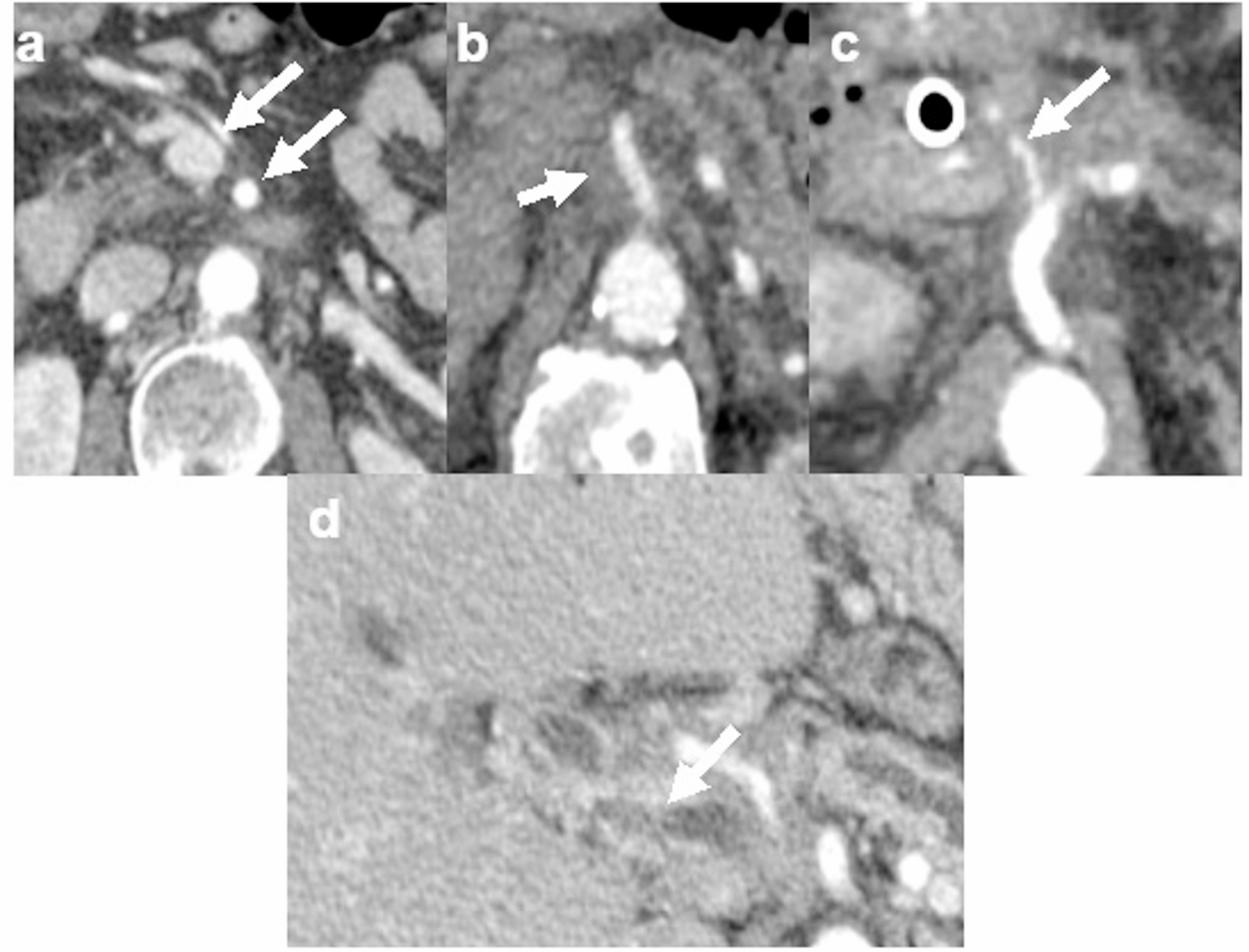
It has been reported that in IVF patients, total OV is positively correlated with the number of oocytes retrieved, fertilization rate, and pregnancy rate. Additionally, the ovarian stromal flow index (FI) has been associated with both oocyte yield and pregnancy outcomes [24]. Although 3D ultrasound is more costly than 2D Doppler ultrasound, it provides valuable insights into ovarian dysfunction, including the assessment of antral follicle count, ovarian and stromal echogenicity, ovarian volume, and blood flow [25]. Currently, the evaluation of ovarian reserve primarily relies on baseline endocrine hormone measurements and ultrasound examination. Ultrasound offers several advantages, such as being radiation-free, non-invasive, and highly reproducible. While conventional 2D ultrasound is effective in detecting pregnancy, it may lack the precision needed to identify subtle abnormalities in the reproductive system. In contrast, 3D transvaginal ultrasound allows for more accurate localization and diagnosis of lesions. The SonoAVC (Sonography-based Automated Volume Count) software used in 3D ultrasound can automatically quantify the number, size, and volume of antral follicles, with each follicle color-coded to prevent measurement duplication. Compared to 2D ultrasound, 3D SonoAVC provides more accurate results in less time (Mindray, Shenzhen, China, model NuewaR9, probe model DE10-3WU, frequency range: 3–12 MHz) (Fig. 2).
Fig. 2
Three-dimensional automated follicle counting in a 39-year-old female patient. The 3D ultrasound reconstruction demonstrates automated identification, numbering, and volumetric measurement of ovarian follicles. The multiplanar views display the spatial distribution of follicles, while the rendered 3D image (bottom right) provides a color-coded visualization of follicular size and morphology, allowing rapid and accurate assessment of follicle count, size, and arrangement
Three-dimensional power Doppler ultrasound, combined with Virtual Organ Computer-aided Analysis (VOCAL) software, provides more precise measurements of OV. The VOCAL technique uses 3D reconstruction to automatically calculate the VI, FI, and VFI, enabling intuitive three-dimensional visualization of the ovarian vascular network and objective quantification of ovarian blood perfusion. Color Doppler imaging, independent of insonation angle, aids in detecting small vessels and low-velocity blood flow, allowing for a more comprehensive evaluation of the ovarian blood supply. In recent years, transvaginal 3D ultrasound has gained increasing attention for its role in diagnosing anovulatory infertility [14, 26].
Studies have suggested that in IVF patients, total OV is positively correlated with the number of oocytes retrieved, fertilization rate, and pregnancy rate. Additionally, the ovarian stromal FI has been linked to both oocyte yield and pregnancy outcomes [24]. 3D ultrasound is particularly useful for guiding individualized treatment in women with a low number of antral follicles, small ovarian volume, and reduced ovarian vascularization. In such cases, treatment may involve a higher dose of gonadotropins and a prolonged stimulation protocol [27]. Although 3D ultrasound is more costly than 2D Doppler ultrasound, it plays a significant role in the comprehensive assessment of ovarian dysfunction, including evaluation of antral follicle count, ovarian and stromal echogenicity, ovarian volume, and blood flow. Therefore, 3D ultrasound provides considerable advantages in optimizing treatment strategies and improving outcomes in IVF patients [25].
Applications of shear wave elastography in ovarian fibrotic disorders: insights from PCOS and POISWE is an ultrasound-based imaging technique that quantitatively assesses tissue stiffness with relatively low operator dependency. It uses a focused acoustic radiation force impulse (ARFI) generated by the ultrasound probe to induce shear waves within the target tissue [28]. These shear waves propagate perpendicularly to the direction of the initial push pulse, with their velocity increasing in stiffer tissues [29]. Ultrafast imaging from the same ultrasound probe is then employed to track the propagation of these shear waves, providing a reliable measure of tissue elasticity [30]. SWE can serve as a noninvasive tool to monitor disease progression, offer quantitative indicators of fertility potential, and assess the effectiveness of clinical interventions aimed at preserving ovarian reserve [31]. It is particularly useful for monitoring ovarian fibrosis and evaluating the potential of therapeutic strategies to maintain ovarian function and prevent fibrotic changes during treatment. It should be noted that different SWE techniques may produce different absolute stiffness values. Point shear wave elastography (pSWE), also referred to as ElastPQ or ARFI-based methods, provides measurements of shear wave velocity or elasticity from a single point or a small region of interest, yielding an averaged value without spatial mapping of tissue stiffness. In contrast, two-dimensional SWE generates a color-coded elastogram within a defined sampling box, enabling visualization of the spatial distribution and heterogeneity of tissue stiffness while providing both qualitative and quantitative information. These technical distinctions are particularly relevant when interpreting differences between transabdominal and transvaginal studies and may partly explain the variability in reported ovarian stiffness measurements across different studies.
PCOS is characterized by increased collagen deposition and ovarian fibrosis, which may contribute to ovarian dysfunction [32]. In an early pilot trial involving 37 PCOS patients and 16 controls, no significant difference in ovarian stiffness was observed using transabdominal SWE [33]. However, subsequent studies consistently reported increased ovarian stiffness in PCOS patients when measured via transvaginal ultrasound, suggesting improved sensitivity and accuracy of this method (Fig. 3).
Fig. 3
Comparison of ovarian 2D shear wave elastography (SWE) findings between a normal ovary and a polycystic ovary. A A 33-year-old healthy female undergoing transvaginal ovarian SWE. The measured elastic modulus values (in kPa) were: Mean 9.23, Max 19.27, Min 0.32. B A 27-year-old female patient with polycystic ovary syndrome (PCOS) underwent transvaginal ovarian SWE. The measured elastic modulus values (in kPa) were: Mean 21.35, Max 34.61, Min 7.60
Compared to normal ovaries, those in PCOS patients tend to have a significantly higher elastic modulus, suggesting increased stromal stiffness, which may reflect ovarian fibrosis. Histological studies have shown that PCOS is associated with an increased antral follicle count, thickening of the tunica albuginea and follicular membranes, and cortical hypertrophy, all of which may influence androgen production and contribute to the characteristic sonographic features of polycystic ovaries [34]. Structural and functional changes in granulosa and theca cells further disrupt folliculogenesis [35].Ovarian stromal stiffness in PCOS is thought to result from follicular cell proliferation and stromal fibrosis, as also demonstrated in animal models of ovarian fibrosis [20]. SWE has emerged as a useful non-invasive tool for assessing ovarian stiffness in PCOS patients. Studies have shown significantly higher elasticity values in PCOS ovaries compared to non-PCOS controls [36]. Increased ovarian stromal volume and thickness in PCOS have been linked to elevated serum testosterone levels and reduced ovarian elasticity [37]. Therefore, SWE-based assessment of ovarian stiffness may serve as a convenient surrogate marker for predicting serum testosterone levels, potentially assisting in the clinical management of PCOS.
There is increasing evidence from both human ovarian tissue and preclinical animal models that individuals with POI exhibit increased ovarian fibrosis, which may contribute to the early decline in ovarian reserve [38]. POI is a heterogeneous condition with iatrogenic, environmental, genetic, and idiopathic causes, characterized by the premature decline of ovarian function [39]. Iatrogenic POI, often resulting from chemotherapy or radiotherapy, is associated with accelerated follicular depletion and early menopause. Interestingly, increased ovarian stiffness and fibrosis have been identified as potential long-term consequences of these treatments, possibly accelerating ovarian aging and impairing future reproductive potential [40].
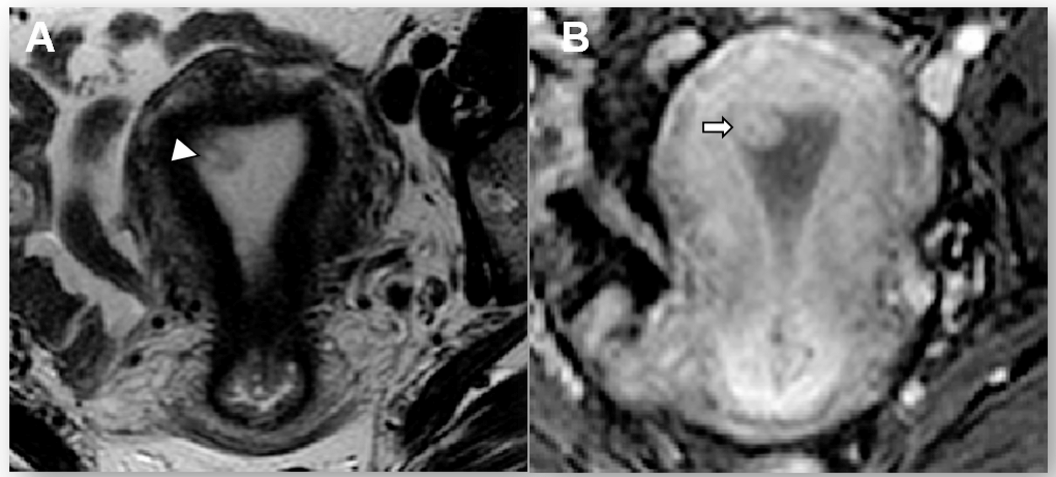
Compared with normal ovaries, those in patients with Hashimoto’s thyroiditis tend to exhibit a higher elastic modulus, suggesting increased ovarian stiffness, which may be indicative of autoimmune-related stromal fibrosis or chronic inflammation (Fig. 4). The rate of fertility decline in POI varies considerably between individuals, making it challenging to predict reproductive outcomes [31]. In this context, SWE may provide a promising non-invasive modality for monitoring disease progression and quantitatively assessing fertility potential in patients with POI. However, SWE is currently limited by small sample sizes, methodological inconsistencies, and the lack of standardized imaging protocols or validated cutoff values. Therefore, further validation through large-scale, prospective studies is needed before SWE can be routinely incorporated into fertility-preservation strategies for patients at risk of treatment-induced POI.
Fig. 4
Comparison of ovarian 2D shear wave elastography (SWE) between a healthy female and a patient with Hashimoto’s thyroiditis. A A 32-year-old healthy female underwent transvaginal ovarian SWE. The mean elastic modulus (E, in kPa) was 8.87, with a maximum of 14.39 and a minimum of 3.70. B A 39-year-old female patient with Hashimoto’s thyroiditis underwent transvaginal ovarian SWE. The mean elastic modulus (E, in kPa) was 21.30, with a maximum of 34.67 and a minimum of 7.05
Advances in CEUS for the evaluation of ovarian diseaseCEUS has gained significant use in reproductive imaging in both human and veterinary medicine. Growing evidence supports its role as a non-invasive and reliable modality for evaluating a range of reproductive disorders [41]. Regarding ovarian function assessment, CEUS enables noninvasive, real-time evaluation of ovarian vascular function during controlled ovarian stimulation. In 2017, Bishop et al. performed CEUS before and 6–8 days after HCG administration (mid-luteal phase) in rhesus monkeys to assess ovarian vascular function (blood volume and vascular flow) during controlled ovarian stimulation. The results showed that CEUS could effectively detect increased blood flow and vascular volume associated with ovarian hyperstimulation syndrome and demonstrate the therapeutic effects of Vascular Endothelial Growth Factor A neutralization. Compared to dynamic contrast-enhanced magnetic resonance imaging, CEUS provides a practical and repeatable method for monitoring ovarian perfusion changes, supporting its clinical relevance. In 2022, Nogueira et al. assessed ovarian vascularization in eight healthy bitches during postovulatory estrus using CEUS. The results indicated an increase in blood flow and pixel intensity as the cycle progressed, with significant increases in perfusion parameters during the luteal phase. Therefore, CEUS proved effective in assessing corpus luteum development, highlighting its utility in monitoring ovarian changes throughout the estrous cycle in dogs [42]. However, studies appear to be limited to animal models, with no clinical research yet available.
Superb microvascular imaging (SMI) is a newer Doppler ultrasound technique that uses an advanced clutter filter to reduce artifacts from breathing and movement, while preserving low-speed blood signals in microvessels. One significant advantage of SMI is its ability to detect slow blood flow signals in microvessels, providing clinicians with more detailed information about blood flow distribution in the target area [43]. Ayaz et al. evaluated ovarian vascularity in 121 girls (ages 3–18) using grayscale ultrasound and five CEUS techniques, including SMI. Color SMI showed the highest interrater agreement (κ = 0.636). The effectiveness of vascular detection was ranked as: monochrome SMI > color SMI > power Doppler > color Doppler > advanced dynamic flow. The study concluded that SMI outperforms conventional Doppler methods and is a promising tool for assessing ovarian vascularity in children [44]. Senyuva et al. conducted a retrospective study evaluating 83 ovaries in 42 women with PCOS using four transabdominal Doppler techniques. The results showed that color and monochrome SMI were significantly more effective than conventional color and power Doppler (p < 0.001) in detecting ovarian stromal vascularity. A stellate vascular pattern was observed in 20.5% of ovaries [45]. Overall, SMI outperforms conventional CEUS techniques for assessing ovarian blood flow in PCOS cases.
The technologies discussed above are expected to play a key role in enhancing the clinical management and quality of life for women affected by declining ovarian function. Table 1 summarizes the main ultrasound imaging modalities and their clinical applications in ovarian reserve assessment, focusing on various techniques used to evaluate ovarian morphology, vascularity, and tissue stiffness. These imaging modalities include 2D and 3D ultrasound, Doppler ultrasound, SWE, CEUS, and AI-assisted imaging, which provide valuable insights into ovarian function and enable more precise, non-invasive assessments. As these technologies continue to advance, they may support more personalized and effective fertility management, offering new opportunities for the early detection and timely intervention of women at risk of ovarian dysfunction.
Contribution of artificial intelligence-driven ultrasound imaging in autoimmune and thyroid-related mechanisms in ovarian dysfunctionAI in medicine refers to knowledge-based and/or data-driven computer systems used to support the prevention, diagnosis, and treatment of diseases [46]. Medical imaging is one of the most prominent areas for AI application in healthcare. In clinical practice, AI in medical imaging is commonly employed for tasks such as image segmentation (identifying and delineating regions of interest), feature extraction (e.g., morphological and textural characteristics), and the development of classification systems for disease diagnosis. With advancements in technologies and algorithms, AI-assisted ultrasound has become an increasingly active area of research. In reproductive medicine, AI-driven ultrasound is primarily applied to automated follicle analysis and ovulation monitoring. As early as 1997, Potocnik et al. applied an optimal thresholding method for the preliminary estimation of ovarian structures and follicle identification [47]. However, limitations such as image noise, challenges in delineating individual follicle boundaries, and insufficient processing speed hindered its use in real-time clinical settings. In subsequent studies, such as that by Faghih et al., follicle detection was achieved by employing various segmentation techniques based on image characteristics (e.g., pixel intensity) and region-specific features (e.g., circularity of the follicular area) [48]. Texture analysis of ultrasound images has also been shown to predict physiological changes related to ovulation, aiding in the optimization of oocyte retrieval timing [49]. When combined with diagnostic classifiers, these image processing methods have shown potential in diagnosing reproductive disorders such as PCOS and POI [50].
Compared to 2D ultrasound, automatic follicle diameter measurement using 3D ultrasound offers several advantages. First, 3D ultrasound data can be stored for detailed post-examination analysis, reducing real-time scan duration. Second, it minimizes operator dependency and subjectivity, improving reproducibility and reducing inter-observer variability [51]. To improve follicular detection, one study proposed an algorithm based on multiple concentric layers for detecting bovine follicles, showing superior performance compared to edge-based methods [52]. Then, an integrated system that combines image denoising, edge detection, and 3D reconstruction algorithms. Their findings suggested that such systems could improve monitoring accuracy and potentially enhance pregnancy outcomes. [
Comments (0)