
Remember me


A 54-year-old male patient was admitted to the Department of General Surgery of The First Hospital of Lanzhou University in 2022. The main complaints were abdominal pain, distension, jaundice (manifestation of skin and sclera yellowing) and pruritus (skin itching) for more than 1 month. Laboratory tests revealed a significant elevation in the serum tumor marker CA19-9, and liver function tests indicated obstructive jaundice (Fig. 1A). Imaging studies (abdominal MRI and CT) revealed a space-occupying lesion in the distal common bile duct, which was highly suspicious for malignancy (Fig. 1B). The patient had a 10-year history of type 2 diabetes mellitus and had previously undergone bladder tumor resection. There was no relevant family history of genetic diseases. After multidisciplinary consultation, the patient underwent pancreaticoduodenectomy. During surgery, white, firm tumor tissue was observed at the distal extremity of the common bile duct (Fig. 1C). Postoperative pathological examination confirmed moderately to poorly differentiated CCA (AJCC-pTNM staging: T2N0Mx). The histological features were characterized by tumor cells arranged in cord-like or glandular patterns infiltrating within a desmoplastic collagenous stroma. The tumor cells exhibited cuboidal to columnar morphology with significant nuclear atypia and a high nuclear‒cytoplasmic ratio. Pathological mitotic figures and perineural invasion were observed. The immunohistochemical staining results were as follows: CK19 (+), TP53 (+), and the Ki-67 proliferation index was 30–40%. Postoperative adjuvant chemotherapy with gemcitabine plus capecitabine was administered, and the patient recovered well. This study was approved by the Medical Ethics Committee of The First Hospital of Lanzhou University (Approval No. LDYYLL2025-954), and written informed consent was obtained from the patients.
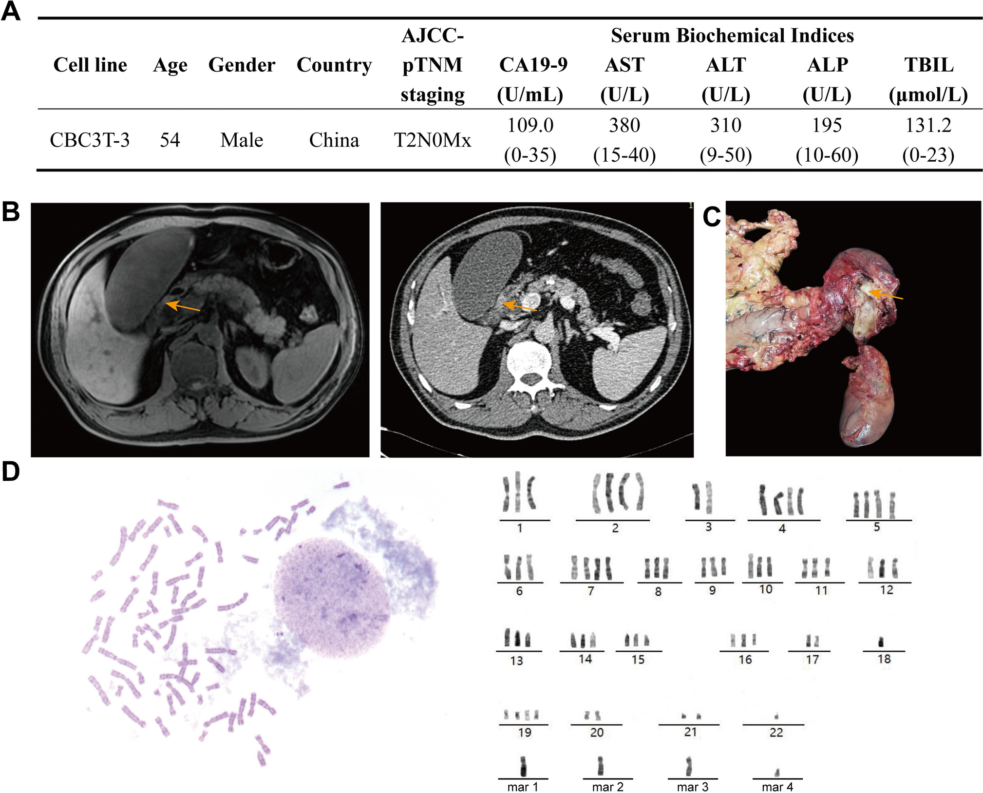
Fig. 1 The alternative text for this image may have been generated using AI.
Clinical characteristics of the patient and identification of CBC3T-3 cells. A Clinical data of the patient. B Preoperative MRI and CT findings suggestive of distal cholangiocarcinoma. C Photograph of the surgical specimen obtained after pancreaticoduodenectomy, showing a white, firm tissue mass protruding into the lumen within the pancreatic segment of the common bile duct. D Chromosome karyotype analysis of CBC3T-3 cells
CBC3T-3 cell line establishmentFresh sterile primary CCA tissue obtained during surgery was immediately placed in precooled DMEM/F-12 medium (Gibco, USA) containing 10% penicillin‒streptomycin dual antibiotics to maintain tissue viability. The tissue was rinsed three times with PBS containing 10% penicillin‒streptomycin dual antibiotics. Using sterile forceps, the fibrous capsule, adipose tissue, and visible blood vessels were carefully removed. The tissue was then minced into small pieces of 1–2 mm3 and transferred into DMEM/F-12 medium containing 200 U of collagenase II for enzymatic digestion for 10 min to obtain a single-cell suspension. Single-cell suspensions were seeded evenly into 6-well plates. After 48 h, the culture medium was replaced, and the cell growth status was observed. Fibroblasts were removed via mechanical scraping. When the cell confluence reached approximately 80%, the tumor cells were further isolated and purified via differential digestion.
Cell cultureThe human extrahepatic CCA cell line TFK-1 was procured from the National Biomedical Experimental Cell Resources Bank (Beijing, China). For cultivation, the cells were grown in RPMI 1640 medium (BI, Israel) containing 10% fetal bovine serum (ABW, USA) and 1% penicillin‒streptomycin solution (BI, Israel). The cells were incubated under standard conditions at 37 °C in a humidified incubator with 5% CO2.
Short tandem repeat (STR) analysisSTR profiling was performed by the China Center for Type Culture Collection (CCTCC; Wuhan, China). In accordance with the ANSI/ATCC ASN-0002–2021 standard, a minimum of 13 STR loci were required for cell identity verification. To genotype CBC3T-3, 21 allele-specific STRs, such as CSF1PO, D5S818, D19S433, etc., were used in the STR multiplex reaction. The STR amplifications were resolved on an Applied Biosystems 3730XL Genetic Analyzer. The resulting STR profile was then mapped against international databases such as ATCC, DSMZ, and CELLOSAURUS to evaluate the degree of matching of this cell line with known STR profiles.
Chromosome karyotype analysisThe cells were grown until the logarithmic growth phase and then incubated with colchicine at a final concentration of 0.2 μg/mL for 2 h to inhibit spindle formation and induce chromosome condensation. After treatment, the cells were harvested and lysed in 0.075 M KCl solution for hypotonic shock, which induced swelling for the dispersion of chromosomes. The cells were then fixed with freshly prepared methanol–glacial acetic acid fixative (3:1, v/v) to preserve the cellular morphology and remove residual hypotonic solution. The fixed cell suspension was dropped onto clean glass slides and dried at 70 °C for 2 h. Chromosomes were stained via Giemsa staining, observed under an optical microscope, and imaged for karyotype analysis.
Cell growth curveThe 96-well plates were seeded with CBT3T-3 cells at a density of 5 × 10³ cells per well. At 6 h, and on days 1, 2, 3, and 4 after seeding, CCK-8 reagent (APExBio, USA) was added to the cells, followed by incubation in the dark for 2 h. The optical density (OD) was then measured at a wavelength of 450 nm. The data were statistically analyzed by using GraphPad Prism 8.0 software (GraphPad Software, USA), and a cell growth curve was plotted to evaluate the proliferation kinetics.
Cell migration and invasionTranswell inserts (8 μm pore size, Corning, USA) were precoated with 50 μL of Matrigel matrix (R&D Systems, USA), which was diluted at a 1:8 ratio with serum-free medium for 30 min at 37 °C for polymerization. After the uncoated medium was removed, 200 μL of a serum-free cell suspension containing 8 × 10⁴ cells was added to the upper chamber, while the lower chamber was filled with 700 μL of medium supplemented with 20% fetal bovine serum (Cell-Box, China) to establish a chemotaxis model. The system was cultured at 37 °C under 5% CO₂ for 48 h, and the cell status was monitored every 12 h. The culture was completed after the set time, after which the cells were fixed in 4% paraformaldehyde for 30 min and stained with 0.1% crystal violet for another 30 min. Images of three arbitrarily selected nonoverlapping fields were recorded via an inverted microscope. The invasive area was quantified via threshold analysis via ImageJ software, and we performed a statistical analysis via GraphPad Prism 8.0.
Transmission electron microscopyThe cells were fixed with electron microscopy fixative (Servicebio, G1102, China) for 2–4 h, followed by three washes with 0.1 mol/L phosphate buffer. The samples embedded in 1% agarose were subjected to postfixation with 1% osmium tetroxide in 0.1 mol/L phosphate buffer. The fixation process lasted for 2 h at room temperature in the dark, after which the samples were thoroughly rinsed three times with the same buffer. After dehydration through a graded ethanol series, the samples were infiltrated with resin and polymerized. Ultrathin sections were obtained via a Leica UC7 (Germany) ultramicrotome, which were then subjected to staining with 2% uranyl acetate in saturated alcohol solution for 8 min in the dark and subsequently dried overnight. Final observation and imaging were conducted using a HITACHI HT7700 (Japan) transmission electron microscope at 80 kV.
Scanning electron microscopyThe cells were fixed with electron microscopy fixative (Servicebio, G1102, China) for 2 h and then rinsed 3 times with 0.1 mol/L phosphate buffer. The samples were dehydrated via a stepwise ethanol gradient and dried via a cold dryer. A small amount of conductive glue was applied to the sample stage, and high-resolution cold-field emission scanning electron microscopy (HITACHI, SU8100, Japan) was used for image observation and acquisition.
Drug sensitivity analysisThe 96-well plates were seeded with CBT3T-3 cells or TFK-1 cells at a density of 5 × 10³ cells per well. After 24 h of culture, different concentrations of gemcitabine, oxaliplatin, cisplatin, fluorouracil or paclitaxel were added for treatment. After 48 h of drug intervention, the original culture medium was discarded, and 200 μL of CCK-8 reagent was added to each well. The plates were incubated at 37 °C for another 2 h. The absorbance values of each well were detected at a wavelength of 450 nm via a microplate reader (BioTek Synergy H1, USA). The experimental data were statistically analyzed and graphed via GraphPad Prism 8.0 software.
Cell line-derived xenograftsFour-week-old NOD-SCID mice were inoculated subcutaneously in the dorsal flank with 5 × 10⁶ CBC3T-3 cells in a 100 μL 1:1 PBS–Matrigel suspension. The animals were monitored every 4 days post-injection. Once the tumors developed, the longest (L) and shortest (W) diameters were measured via a Vernier caliper, and the tumor volume was calculated. At 10 weeks post-injection, the mice were euthanized, the subcutaneous tumors were excised for imaging and weighing, and portions of the tumor tissue were fixed for hematoxylin–eosin (HE) and immunohistochemical staining. The remaining tissues were stored at – 80 °C.
H&E and immunohistochemical stainingFor immunohistochemistry, formalin-fixed, paraffin-embedded tissues were sectioned and processed through deparaffinization, rehydration, antigen retrieval, and serum blocking. The sections were then incubated overnight at 4 °C with primary antibodies against Ki-67 (1:500, GB111499, Servicebio), TP53 (1:1000, GB12626, Servicebio), and CK19 (1:500, GB11197, Servicebio). This was followed by washing with PBS, incubation with species-matched secondary antibodies, and DAB chromogenic development. Hematoxylin was used for nuclear counterstaining, and the stained sections were visualized under a Nikon (Japan) microscope.
WESLibraries for exome sequencing were constructed from genomic DNA isolated from the patient's adjacent normal tissue and CBC3T-3 cells. The libraries were subjected to exome capture via the Agilent SureSelect Human All Exon V6 system (Novogene, Beijing, China) and sequenced on an Illumina NovaSeq 6000 platform. The generated raw data were aligned via BWA and interrogated via the GATK pipeline for variant calling. The spectrum of final annotated variants included single nucleotide polymorphisms (SNPs), insertions and deletions (InDels), and copy number variations (CNVs).
Statistical analysisThe experiments in this research were performed in three biological replicates (n = 3). An unpaired two-tailed Student's t test was used to assess the significant differences between the compared groups. All the statistical analyses were performed via GraphPad Prism 8, and P < 0.05 was considered statistically significant.
Comments (0)