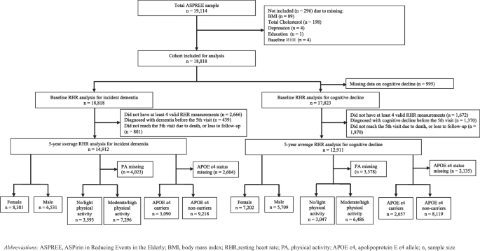
In this secondary analysis of a large prospective cohort study, we explored the association of RHR with cognitive decline and incident dementia in older adults. Participants with a baseline RHR ≥ 80 bpm exhibited a significantly higher risk of cognitive decline compared to those with an RHR of 60–69 bpm. Similar patterns emerged for the 5-year average RHR, where RHR levels of 70–79 bpm and ≥ 80 bpm were both significantly associated with an increased risk of cognitive decline. Subgroup analyses yielded broadly consistent trends, though some associations did not reach statistical significance, likely due to smaller sample sizes. Notably, sex-stratified analysis revealed distinct patterns, among females, HRs tended to increase progressively across higher RHR categories, whereas the pattern among males was less consistent. When stratified by physical activity, elevated RHR was associated with a higher risk of cognitive decline among participants reporting moderate-to-high physical activity, but not among those reporting no or light physical activity. Crucially, within this healthy cohort, no significant associations were observed between RHR and incident dementia.
Our findings regarding cognitive decline align with two previous longitudinal studies [13, 18]. Imahori et al. examined 2,147 participants (≥ 60 years, mean age 70.6 years) from the Swedish National Study on Aging and Care in Kungsholmen (SNAC-K) cohort, reporting that baseline RHRs of 70–79 bpm and ≥ 80 bpm were associated with greater cognitive decline over 11.4 years of follow-up compared to 60–69 bpm [18]. Similarly, Deng et al. investigated 339,901 participants aged 46–69 years (mean age 57.3 years) from the UK Biobank, finding that higher baseline RHRs (70–79 bpm and ≥ 80 bpm) were significantly associated with slower reaction times over an average follow-up of approximately 8.5 years [13]. Conversely, Haring et al., utilized data from the Women’s Health Initiative Memory Study (WHIMS) involving postmenopausal women (≥ 63 years, mean age 69 years) and found no significant association between a 9-year average RHR and cognitive impairment [14]. This discrepancy may partly reflect differences in RHR measurement and categorisation; Haring et al., utilized manual pulse assessments and operationalized RHR using tertiles (51–66, 67–70, and 71–92 bpm) with the lowest group as the reference [14]. It is worth noting that an RHR < 60 bpm may reflect conduction abnormalities or reduced cerebral perfusion, potentially biasing associations with cognitive decline when used indiscriminately within a reference group [18, 37].
For incident dementia, our null findings contrast with those of Imahori et al. and Deng et al., both of whom reported an increased dementia risk among participants with RHR ≥ 80 bpm compared with 60–69 bpm [13, 18]. Several methodological variations may explain these discrepancies. Unlike previous studies relying on a single baseline RHR measurement [13, 18], our baseline RHR was derived from the average of three distinct measurements, providing a more stable clinical estimate of long-term exposure. Differences in participant baseline characteristics are also highly relevant. The ASPREE cohort comprised community-dwelling older adults free of CVD and physical disability at baseline. In contrast, the SNAC-K cohort included participants from institutional settings, such as nursing homes and special accommodation facilities, who generally exhibited more severe functional and cognitive impairments [38]. Additionally, variations in CVD screening protocols and dementia case ascertainment methods across cohorts may have contributed to divergent findings.
Collectively, these findings suggest that elevated RHR may be more strongly associated with early physiological ageing and cognitive changes than with the later clinical manifestations of overt neurodegenerative disease. The observed association with cognitive decline, but not dementia, supports the hypothesis that RHR functions as a systemic biomarker of subclinical autonomic or cardiovascular dysregulation rather than primary neurodegeneration. Because cognitive decline frequently precedes the onset of dementia by many years [39], it represents a more sensitive readout of early homeostatic dysregulation.
Furthermore, the magnitude of the association between RHR and cognitive decline in the present study was modest relative to established risk factors, including advancing age, cardiovascular risk burden, and APOE genotype [40,41,42]. This indicates that while RHR captures critical aspects of underlying biological vulnerability, it is unlikely to be a major standalone determinant of dementia risk. Mechanistically, an elevated RHR is an established indicator of autonomic imbalance, metabolic stress, and systemic inflammation, all of which may contribute to vascular and cognitive aging [43, 44]. However, clinical progression to dementia likely requires the accumulation of additional neuropathological changes that are not directly reflected by RHR alterations alone. These data therefore position RHR as a potential early indicator of functional decline rather than as a specific predictor of dementia.
Several biological pathways may help contextualize the observed association between elevated RHR and cognitive decline, although these mechanisms remain speculative. An elevated RHR may reflect altered autonomic regulation through the central autonomic network, which integrates cortical, limbic, and brainstem regions involved in both heart rate control and cognitive processes [45,46,47]. More broadly, RHR can serve as a multidimensional marker of biological aging and organism-level homeostasis, capturing cumulative cardiovascular load as well as metabolic, inflammatory, autonomic, and vascular dysregulation [44, 48]. Rather than acting through a single pathway, an elevated RHR may capture systemic physiological dysregulation that accompanies aging, including brain aging [49], the promotion of adverse cardiovascular remodelling [50, 51], and the disruption of autonomic balance [11]. Through these concurrent pathways, an elevated RHR may signal compromised vascular health, impaired cerebral perfusion, or altered neurovascular coupling, all of which can negatively impact cognitive performance.
The stronger associations observed among females in our study reflects sex-specific differences in autonomic regulation, cardiovascular physiology, or vulnerability to age-related physiological changes. Meanwhile, the apparent interaction with physical activity should be interpreted cautiously, as it may reflect underlying differences in health status or residual confounding rather than a true modifying effect. Overall, these findings suggest that an elevated RHR serves as a signature of broader physiological dysregulation related to cognitive decline, rather than acting via a single causal pathway.
The clinical implications of these findings should be interpreted with caution. Although the associations between an elevated RHR and cognitive decline were statistically significant in this large cohort, the effect sizes were small. Given this modest effect size, the incremental contribution of RHR to model discrimination is likely limited. RHR is therefore unlikely to function as a strong standalone predictor, but it may provide valuable complementary information alongside established risk factors. Importantly, RHR is a simple, non-invasive, and universally available metric, which enhances its utility at a population level. Combining RHR measurements with standardised tools, such as the MMSE [52] or Montreal Cognitive Assessment [53], may help identify individuals experiencing early cognitive ageing who would benefit from closer monitoring or preventive interventions, such as physical activity, dietary adjustments, and vascular risk control, before irreversible neurodegenerative damage occurs [7, 36]. Future research should formally evaluate whether incorporating RHR improves predictive performance metrics, such as the C-statistic or net reclassification improvement (NRI), in established dementia risk models.
Strengths and limitations
This study has several notable strengths. The large sample size, longitudinal design, and rigorous outcome ascertainment strengthen the reliability of our findings. By examining both baseline RHR and a 5-year longitudinal average, we provided a more stable estimate of long-term exposure than studies utilizing single-timepoint measurements. To our knowledge, this is the first study to evaluate both baseline and longitudinally averaged RHR in relation to cognitive decline and dementia within the same cohort.
Nonetheless, several limitations must be acknowledged. The ASPREE cohort comprised relatively healthy older adults free of CVD and cognitive impairment at baseline, which may limit the generalizability of our findings to broader, multi-morbid ageing populations and could have attenuated the observed associations. Additionally, participants were not required to fast or refrain from caffeine or smoking prior to clinical measurements, which might induce short-term physiological fluctuations. However, this concern was mitigated because RHR was measured three times and averaged at each clinical visit, and further minimized by our use of the 5-year average RHR. While major confounders were adjusted for in our models, residual confounding cannot be entirely excluded. Unmeasured or incompletely measured factors, such as daily physical activity levels, sleep quality, subclinical CVD, and longitudinal changes in medication regimens, may concurrently influence both RHR and cognitive outcomes. In addition, reverse causation remains a possibility, as early neurodegenerative changes can disrupt central autonomic regulation and consequently alter RHR. Although the use of 5-year average RHR provides a more stable estimate of long-term exposure, it does not fully eliminate these temporal concerns.



Comments (0)