
Remember me



Primary immunodeficiency (PID) is a group of genetic disorders characterized by immune dysfunction, some of which may present with autoimmune manifestations that mimic connective tissue diseases (CTD). While most cases manifest during childhood, there are instances of adult-onset diagnoses. Undiagnosed PID patients may be diagnosed as adults with atypical CTD, leading to resistance to conventional treatment or susceptibility to infection. In this case, a patient initially diagnosed with rheumatoid arthritis (RA) who also presented with multiple endocrine disorders and clinical features resembling those of systemic lupus erythematosus (SLE) was ultimately diagnosed with lipopolysaccharide-responsive beige-like anchor protein (LRBA) deficiency.
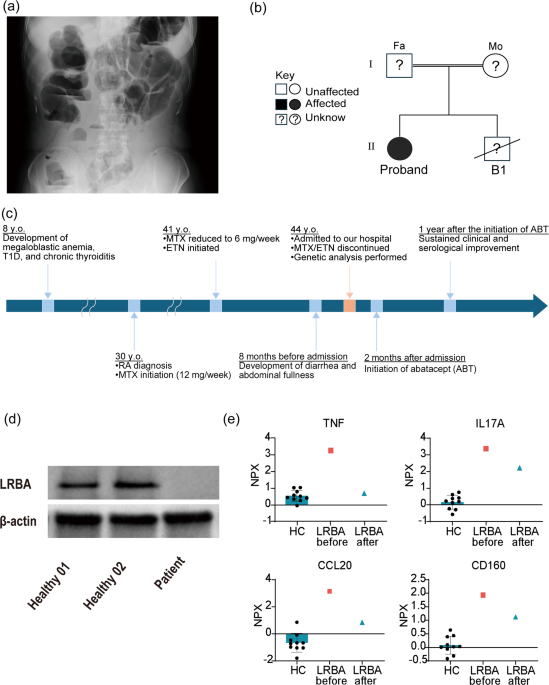
A 44-year-old Japanese woman with a 14-year history of seronegative RA was treated with methotrexate (MTX) at 12 mg/week. Gastrointestinal symptoms developed three years prior to admission, leading to a reduction in MTX dosage to 6 mg/week. Subsequently, the patient’s arthritis gradually worsened, and etanercept, a tumor necrosis factor (TNF) receptor inhibitor, was initiated four months before admission. Joint destruction was not observed despite the prolonged disease duration of RA. The patient had experienced persistent diarrhea and abdominal fullness for more than 8 months prior to admission. Upon admission, the patient presented with weight loss and severe gastrointestinal symptoms, including diarrhea and abdominal pain. At that time, the patient also presented with pancytopenia due to megaloblastic anemia, hypocomplementemia, hypogammaglobulinemia (Table S1), and severe intestinal pseudo-obstruction (Fig. 1a). Her medical history included megaloblastic anemia, esophageal candidiasis, type 1 diabetes, and chronic thyroiditis, starting from the age of 8. The underlying cause of megaloblastic anemia had been investigated during childhood; however, both anti-parietal cell and anti-intrinsic factor antibody levels were within normal limits, direct antiglobulin test was negative, and no definitive cause was identified. Type 1 diabetes was treated with insulin and chronic thyroiditis with thyroxine. Early-onset endocrinopathies and esophageal candidiasis suggested autoimmune polyendocrine syndrome, but no definitive diagnosis was made. Notably, her brother died of unexplained severe diarrhea during childhood, and this family history raised the possibility of a congenital autoimmune or endocrine disorder due to an underlying genetic abnormality (Fig. 1b). Upon admission, as possible differential diagnoses, SLE with lupus enteritis was suspected based on the presence of pancytopenia, hypocomplementemia, and chronic arthritis. However, the patient had a low-titer ANA (1:80), and other serological markers for SLE were negative. In addition, the chronic course of symptoms and the presence of early onset endocrinopathies were atypical for SLE. Considering her early-onset endocrine abnormalities, multiple autoimmune manifestations (anemia, arthritis, enteropathy), and family history of her brother’s death, we suspected an underlying PID and performed genetic analysis (Fig. 1c). Genetic analysis identified a heterozygous LRBA splice-site variant (c.1162-1G > A) and a possible heterozygous deletion involving exons 18–41. Western blot analysis demonstrated loss of LRBA protein expression (Fig. 1d; Supplementary Material). These findings were consistent with LRBA deficiency and suggested compound heterozygous LRBA mutations. The deletion breakpoints remain to be determined. Following the diagnosis, she was treated with abatacept (ABT, CTLA4-Ig fusion protein) at a dose of 125 mg subcutaneously weekly, known to be effective against LRBA deficiency [1], which markedly improved her symptoms and laboratory findings within weeks, with resolution of diarrhea and normalization of blood counts and complement levels over the subsequent months. Since ABT initiation, symptoms have not recurred, and laboratory values have remained stable for several years. (Table S2).
Fig. 1
Clinical characteristics, treatment course, and immunological findings of the patient with LRBA deficiency. (a) X-ray of the abdomen (standing position) at administration. It revealed severe intestinal dilatation and multiple air-fluid levels, consistent with findings of ileus. (b) Pedigree of the family. The patient is shown as a filled circle. The slashed symbol indicates a deceased individual. The younger brother died in infancy, and his LRBA status remains unknown. Both parents were unavailable for genetic evaluation. (c) Clinical course of the patient. The patient developed megaloblastic anemia, type 1 diabetes mellitus, and chronic thyroiditis during childhood, followed by rheumatoid arthritis (RA) in adulthood. RA was treated with methotrexate (MTX) and etanercept (ETN); however, diarrhea and abdominal fullness subsequently developed. Genetic analysis performed after admission revealed LRBA deficiency. Abatacept (ABT) treatment resulted in sustained clinical and serological improvement. (d) Western blotting analysis of LRBA protein expression revealed markedly reduced LRBA expression in the patient. (e) Serum levels of inflammation-related proteins in the LRBA-deficient patient were quantified using the proximity extension assay (Olink® Target, 384 proteins). Sera from ten healthy controls (HC) were used as references. Among them, TNF-α, IL-17 A, CCL20, and CD160 showed significant changes before and after treatment. TNF: tumor necrosis factor, IL17A: interleukin 17 A, CCL20: chemokine (C-C motif) ligand 20, NPX: normalized protein expression
To gain insight into the pathogenic mechanisms in this LRBA-deficient case, we measured 384 inflammation-related serum proteins before/after treatment and in ten healthy controls using the proximity extension assay (Olink® Target). After 12 months of treatment, there was a marked improvement in TNF (3.26 to 0.69), interleukin 17 A (IL17A, from 3.36 to 2.22 normalized protein expression), chemokine (C-C motif) ligand 20 (CCL20, 3.14 to 0.84), and CD160 levels (1.93 to 1.12, Fig. 1e).
LRBA deficiency is one of the PIDs associated with autoimmune manifestations. LRBA plays a pivotal role in the intracellular trafficking of cytotoxic T lymphocyte protein-4 (CTLA-4) by re-routing it from lysosomal degradation back to recycling endosomes in activated T cells. In LRBA deficiency, the levels of CTLA-4 on activated T cells decrease due to increased lysosomal degradation, leading to diverse clinical manifestations, including autoimmune manifestations, enteropathy, and endocrinopathy.
In cases where symptoms of LRBA deficiency appear in early childhood, the diagnostic pathway typically leads to suspicion of PID and subsequent genetic testing. However, adult-onset cases, such as the case of this patient, are often atypical for both PID and CTD, and given the limited awareness of LRBA deficiency, reaching a definitive diagnosis may be particularly challenging. The availability of specific clinical markers would greatly facilitate earlier diagnosis, but currently, no such established biomarkers exist. In this case, TNF, IL17A, CCL20, and CD160 levels decreased after ABT treatment.
In LRBA deficiency, an imbalance of CD4+ T cells is reported with high frequencies of Th1, CD4+ IFN-γ+ IL‐17+, and Th22 cells, along with a low frequency of regulatory T (Treg) cells [2]. Overproduction of IL-17 may induce various CTD symptoms, such as arthritis and pancytopenia. CCL20 is associated with lymphocyte induction. In Th17 cells, IL-17 A induces the expression of CCL20 in the small intestine, promoting intestinal migration via the CCR6/CCL20 axis [3]. The CCR6/CCL20 axis can induce enterocolitis and pseudo-obstruction. LRBA is also known to influence type 3 innate lymphoid cells (ILC3), which control the balance of Th17 cells and Treg cells in the large intestine [4]. CD160 expression is linked to CD8+ T cells and is known to be associated with their activity in mouse models [5]. Activated CD8+ T cells may trigger inflammation in the pancreas and thyroid glands, leading to type 1 diabetes and chronic thyroiditis.
This case highlights the importance of considering PIDs such as LRBA deficiency in adult patients presenting with atypical autoimmune manifestations. A previous systematic review described that LRBA deficiency is typically diagnosed during early childhood, with a median age at onset of 1.8 years [1]. In contrast, our patient developed in adulthood with CTD-like manifestations. Our patient had compound heterozygous LRBA variants, which may have been related to the adult diagnosis. The patient had shown various abnormalities, and gastrointestinal symptoms raised suspicion of PID and led to the diagnosis. Gastrointestinal involvement is common in LRBA deficiency, occurring in approximately 68%, and may reflect immune dysregulation involving IL-17 A and CCL20, both of which were elevated in our inflammatory protein analysis [1, 3, 4]. In contrast, articular manifestations are less common, although our patient had longstanding arthritis with elevated TNF levels [1]. These cytokines may represent potential clinical markers of LRBA deficiency, although these findings remain exploratory in this single case. The successful use of abatacept emphasizes the therapeutic benefit of restoring CTLA-4–mediated inhibitory signaling in genetically confirmed LRBA deficiency, rather than acting solely as a conventional immunosuppressive agent. In patients with refractory or atypical autoimmunity, especially with early endocrine or gastrointestinal involvement, clinicians should consider underlying PIDs and perform genetic analysis in appropriate clinical settings.
In conclusion, we present a challenging case of apparent CTD in an adult patient who was ultimately diagnosed with PID due to LRBA deficiency. Patients with PIDs may phenotypically manifest a variety of autoimmune diseases. Since LRBA deficiency is effectively treated with abatacept, it is strongly recommended to consider the possibility of autoimmune diseases associated with PIDs and performing genetic analysis in clinical practice.
Comments (0)