In recent years, clinical entities at the intersection of uveitis and genetics have gained increasing recognition. These include IRDs masquerading as uveitis, IRDs complicated by uveitis, and uveitis of genetic origin [4]. The latter can present with an array of manifestations including conjunctivitis, episcleritis, anterior and posterior uveitis in syndromic contexts, such as ALPK1-related ROSAH syndrome or NOD2-related Blau syndrome [5]. However, to our knowledge, npAIR is not commonly associated with these conditions.
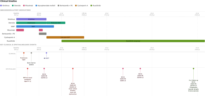
Here, we report the case of a young girl who developed npAIR in the context of a TLR7-related interferonopathy, following an unsuccessful attempt to taper her immunosuppressive therapy for autoimmune thrombocytopenia and hemolytic anemia. Her case had previously been described in the literature by David et al., although without documentation of retinal involvement [1]. After undergoing hematopoietic stem cell transplantation, she developed graft-verus-host disease (GVHD), which was subsequently controlled with long term treatment with the JAK inhibitor Ruxolitinib. During a 3-year follow-up, her retinal findings progressed gradually, with macular thinning and pigmentary changes suggestive of post-inflammatory damage. However, it remains uncertain whether disease stability was linked to immune reconstitution or the continued effect of systemic immunomodulation. Taken together, the immunologic context and timing of symptoms support a possible association between the underlying TLR7-related condition and immune-mediated retinal injury.
Interestingly, the severe outer nuclear layer thinning and slowly progressive retinal degeneration despite immunosuppression bore some resemblance with the recently described entity of acute outer retinopathy (AOR) [6]. AOR is defined by plaque-like yellow-grayish lesions with hyperautofluorescence on FAF and full-length photoreceptor degeneration, typically accompanied by the angular sign of Henle fiber layer hyperreflectivity (ASHH) on OCT [6]. In our case, ASHH was not identified, which may reflect either its true absence or the possibility that this transient feature had already resolved by the time of delayed ophthalmic evaluation. As AOR has been proposed to represent severe forms within the spectrum of autoimmune retinopathy, it cannot be entirely excluded; however, npAIR serves as the most fitting classification in this setting.
Ocular involvement has previously been described in association with TLR7-related disease, particularly in the form of neuromyelitis optica [2]. Moreover, murine models of autoimmune uveitis have demonstrated aberrant upregulation of TLR7 signaling [7], further supporting its potential role in immune-mediated retinal pathology in this case.
Interestingly, npAIR has also been described in association with non-monogenic SLE and other inborn errors of immunity, including common variable immunodeficiency (CVID) [8, 9]. Muzio et al. described a 34-year-old male patient with CVID carrying one pathogenic variant and two VUS in the TACI gene [8]. This patient was diagnosed with npAIR and exhibited multiple hyperautofluorescent annular lesions in the peripapillary area and retinal periphery of the left eye, along with subtle hyperautofluorescent lesions in the right eye. Similarly, Wiley et al. reported a 40-year-old woman with CVID who presented with rapidly progressive bilateral visual field loss, ellipsoid zone constriction, and cystoid maculopathy [9]. Her clinical picture stabilized with mycophenolate mofetil and intravenous immunoglobulin therapy, resulting in preservation of central vision. These cases differ in clinical presentation and imaging, underscoring the marked heterogeneity of npAIR and its nature as a challenging diagnosis of exclusion [10]. In this context, awareness of underlying immune dysregulation, particularly in patients with known genetic or syndromic backgrounds, may aid in recognizing such atypical presentations. Conversely, the role of anti-retinal antibodies remains controversial. Although they can be detected in both paraneoplastic and non-paraneoplastic autoimmune retinopathy, their diagnostic value is limited by variable sensitivity and poor specificity, with reported positivity also in healthy individuals and other retinal diseases. As such, their detection is not required to confirm the diagnosis, particularly when clinical and electrophysiological findings are strongly suggestive [10].
In summary, we present the case of a young girl with TLR7-related autoimmune thrombocytopenia and progressive leukoencephalopathy who developed an atypical form of npAIR. This case broadens the spectrum of clinical manifestations associated with monogenic SLE. While her condition remained stable over time, it is difficult to disentangle the respective contributions of immune reconstitution and ongoing systemic therapy. Nonetheless, this observation reinforces the importance of maintaining immune control in patients with underlying interferonopathies and ocular autoimmunity. In light of this, regular ophthalmological screening may be considered in patients with interferonopathies to allow early detection of potential ocular involvement.




Comments (0)