
Remember me
Particle therapy with protons and carbon ions offers improved dose conformity compared with conventional photon radiotherapy due to the characteristic Bragg peak and reduced exit dose, enabling enhanced sparing of surrounding organs-at-risk (OARs) while maintaining adequate tumour coverage (Shanbhag 2023, Knäusl et al 2024, Liu et al 2024). However, these physical properties make particle therapy highly sensitive to uncertainties in tissue density, patient setup, and anatomical variations (Phillips et al 1992, Pakela et al 2022). Changes in anatomy and target position can significantly affect the delivered dose distribution (Rietzel and Bert 2010, Boria et al 2018). Intrafractional motion is particularly relevant in thoracic and abdominal regions, where respiratory motion constitutes the primary driving factor, whereas interfractional anatomical variations could potentially occur across all treatment sites. This intrafractional motion remains a major challenge in particle therapy (Qubala et al 2024). In proton therapy, motion is primarily a physical dosimetric problem driven by range sensitivity and steep distal dose gradients, making the delivered dose highly susceptible to temporal misalignment between beam delivery and anatomy. In carbon ion therapy, motion has a similar physical impact but additionally induces larger variations in linear energy transfer and relative biological effectiveness, adding a biological component to the overall uncertainty. For affected indications, such as e.g. lung, pancreas or liver, temporal variations in target position and surrounding anatomy may lead to deviations between the planned and delivered dose distributions, potentially degrading target coverage and increasing dose to adjacent OARs (Pakela et al 2022). The problem is further exacerbated when dynamic delivery techniques such as pencil beam scanning (PBS) are used (Bert et al 2008). In PBS treatments, the sequential delivery of discrete beam spots interacts with target motion, producing the so-called interplay effect, which may introduce substantial dose heterogeneities within the target volume (Li et al 2022). These effects are particularly relevant in hypofractionated regimens, where the reduced number of fractions limits the averaging of motion-induced dose variations (Liu et al 2024).
Various motion-management strategies have been proposed to mitigate these intrafractional effects, including four-dimensional (4D) treatment planning using respiratory-correlated imaging, internal target volume (ITV) concepts, rescanning techniques, respiratory gating, breath-hold (BH) strategies, and tumour tracking complemented by adaptive treatment approaches to address interfractional changes (Shirato et al 2000, Keall et al 2006, Czerska et al 2021, Li et al 2022, Knäusl et al 2024, Qubala et al 2024, Belikhin et al 2024). To minimise the effect of interfractional uncertainties, robust optimisation methods are increasingly employed to account for setup and range during treatment planning and evaluation (Ribeiro et al 2021, Boer et al 2022, Rana and Rosenfeld 2022, Spautz et al 2023). Despite these developments, clinical implementation of motion mitigation and 4D dose evaluation remains heterogeneous across institutions, reflecting differences in imaging capabilities, treatment planning systems, delivery technologies, and clinical workflows (Zhang et al 2023, Czerska et al 2025, Fracchiolla et al 2026). Additionally, differences in motion assessment, temporal delivery modelling, and dose accumulation methodologies may lead to significant variability in reported motion effects across institutions.
Previous reports and task group recommendations have reviewed motion management techniques and provided guidance on their implementation and quality assurance in particle therapy (Knopf et al 2014, 2016, Alina et al 2020, Ribeiro et al 2020, Chuong et al 2021, Czerska et al 2021, Knäusl et al 2024). However, rapid technological developments—including advances in imaging, artificial intelligence, treatment delivery systems, and adaptive workflows—continue to evolve in the field. At the same time, the increasing adoption of PBS delivery and hypofractionated treatments has highlighted the need for robust and standardised approaches to evaluate motion-induced dose uncertainties and interplay effects.
This work, initiated by the 4D subgroup of the European Particle Therapy Network (EPTN) workpackage 5 (WP5), involving 20 particle therapy and academic research centres, aimed to provide a comprehensive state-of-the-art overview of clinically applied motion management and 4D treatment planning approaches in particle therapy. It focused on the mitigation of intrafractional movements for thoracic and abdominal indications. The objective of this review was to systematically analyse these clinically applied methodologies covering motion mitigation techniques, robustness evaluation, and interplay assessment, highlighting their strengths, limitations, and areas of clinical variability. Based on this analysis, the study sought to outline insights into clinical practice and methodological considerations to support the development of more consistent and standardised frameworks for 4D dose assessment in clinical routine, ultimately contributing to improved confidence that the planned dose accurately reflects the dose delivered to the patient.
This work was conducted as a systematic review following the Preferred Reporting Items for Systematic Reviews and Meta-Analyses guidelines (Moher et al 2009). A structured literature search was performed to identify studies addressing motion management, 4D treatment planning, and dose evaluation in proton and carbon ion therapy.
Systematic searches were conducted in the PubMed and Web of Science databases to identify relevant publications published between January 2020 and December 2025. The search strategy targeted studies addressing 4D planning approaches, motion mitigation techniques, and motion-aware dose evaluation in particle therapy. Search terms were carefully selected by a dedicated team within the EPTN WP5 4D subgroup (details are listed in the supplementary materials). After the removal of duplicate records, all retrieved publications were independently screened for relevance by two members of the working group. In cases of disagreement during the screening process, a third reviewer served as conflict resolver to make the final decision on inclusion or exclusion of the publication.
A team of eight experts performed the initial assessment of the identified studies. Considering the title and abstract, publications were evaluated for eligibility based on predefined inclusion and exclusion criteria (see supplementary). They were then categorised according to key thematic areas reflecting the main components of 4D motion management workflows in particle therapy as follows: motion management, imaging, treatment planning, clinical indications, phantom studies, and research and future perspectives (table 1). Individual articles could be assigned to more than one subtopic. Relevant publications were then assigned to two or three topic leads per thematic area for detailed evaluation and synthesis.
Table 1. Defined thematic areas and assigned publications. Papers could be assigned to more than one subtopic.
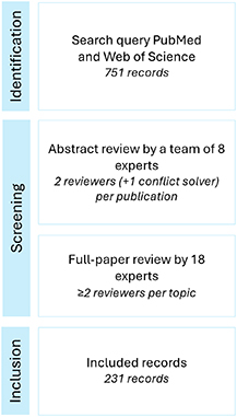
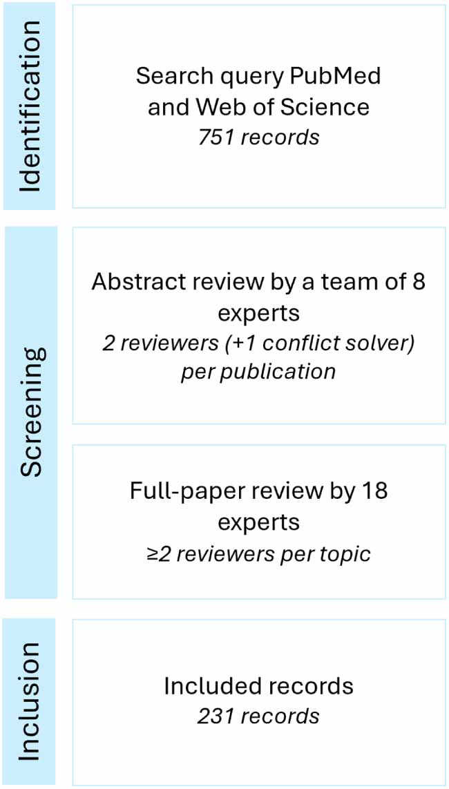
SubtopicAmount of papers includedMotion management Patient-related46Beam-related58Imaging Pre-treatment35Patient setup17Patient monitoring20Dose monitoring21Treatment planning Plan optimisation73Plan evaluation86Clinical indications Lung76Pancreas22Liver35Lymphoma/thymoma8Oesophagus12Breast17Phantom studies38Research and future perspectives40In total, 751 publications were identified. Following the screening and eligibility assessment process, 231 publications were included in the final review (figure 1). Studies not identified through the database search but considered relevant for introduction of a dedicated thematic area were also included, adding another 18 publications.
Figure 1. Overview of the literature screening and publication selection. Searches were conducted in the time period from January 2020 to December 2025.
Download figure:
Standard image High-resolution image 3.1. Limiting the tumour motionPatient-related motion mitigation is fundamentally constrained by what patients can tolerate and where the respiratory motion can be reasonably reproduced during delivery and verification.
Deep inspiration BH (DIBH) and deep expiration BH (DEBH) protocols, as well as related BH techniques, are key strategies when adequate patient cooperation is achievable (Emert et al 2021, Sabouri et al 2024). Multi-centre guidance and surveys have identified BH as one of the most commonly applied active methods in clinical practice. Real-time motion management has been adopted by 85% of surveyed US proton centres, and DIBH/DEBH was reported as the most frequent approach (Zhang et al 2023, Kang et al 2025). BH reduces the irradiated volume and OAR dose, but it prolongs the treatment time depending on the indication and the patient’s capabilities. Its success depends on training, coaching, and tolerance of repeated short BHs (Wang et al 2020, Emert et al 2021, Mah et al 2022). Several different monitoring systems are in clinical use, including spirometry-based BH control systems, infrared marker-based respiratory monitoring systems, and optical surface imaging-based guidance with structured coaching. Reproducibility is high when programs formalise training and verify it with repeated BH computed tomography (CT) or cone-beam CT (CBCT) (Andersson et al 2020, Loap et al 2021, Mah et al 2022, Patel et al 2022, Li et al 2022, Costin and Marcu 2024, Sabouri et al 2024). In a physiology-driven comparison, enhanced DIBH outperformed high-frequency percussive ventilation for motion suppression and was preferred by 80% of participants, indicating that non-intubated strategies with short BHs are generally better tolerated than invasive ventilation paradigms (Emert et al 2021). If BH is not feasible or residual motion persists, abdominal compression can be applied to mechanically limit diaphragmatic excursion and reduce respiratory motion amplitude (Schneider et al 2023). Non-intubated ventilation-based approaches represent an additional option. Continuous positive airway pressure has been reported to reduce heart and lung dose in breast and thoracic settings, particularly when BH is not feasible (Ko et al 2021, Costin and Marcu 2024). Non-invasive high-frequency oscillatory ventilation has also been shown to significantly reduce diaphragmatic motion while remaining tolerable for up to 30 minutes in volunteers (Zhang et al 2022). More invasive techniques, including apneic oxygenation and high-frequency jet or percussive ventilation, can nearly immobilise thoraco-abdominal targets but require anaesthesia support and specialised teams. Case-level carbon ion therapy has demonstrated the feasibility of high-frequency oscillatory ventilation under general anaesthesia for irradiation of multiple lung metastases in a single fraction, although this resource-intensive approach is limited to carefully selected patients (Li et al 2022, Zhang et al 2025).
Audio-visual biofeedback synchronising short BHs with synchrotron flattops improved target coverage and dose homogeneity without prolonging sessions and even reduced treatment time compared with fixed-cycle guidance (He and Li 2021, 2022).
3.2. Modifying beam deliveryBeam-related motion management techniques mitigate motion-induced dose degradation by modifying the temporal, spatial, or logical structure of beam delivery rather than patient anatomy or physiological processes (Ribeiro et al 2020, den Boer et al 2021, Rana and Rosenfeld 2021, Steinsberger et al 2021, Lee et al 2024, Liang et al 2024, Sabouri et al 2024, Kang et al 2025, Liu et al 2024). By that, target coverage can be maintained, and dose to OARs can be reduced.
Among beam-related techniques, rescanning, also referred to as repainting, has been investigated most extensively in clinical settings. It is based on delivering the planned dose multiple times with a fraction of the spot weight per scan. By temporally averaging motion-induced dose heterogeneities, rescanning can improve target dose homogeneity and robustness, particularly in the presence of quasi-periodic respiratory motion (Ribeiro et al 2020, Meijers et al 2020, Rana and Rosenfeld 2021, Worm et al 2021). Several rescanning concepts have been proposed, including layer-based rescanning, volumetric rescanning, and uniform-spaced fast rescanning, which exhibit different sensitivities to motion characteristics and delivery efficiency (Rana and Rosenfeld 2021, Lee et al 2024, Zhang et al 2025).
The effectiveness of rescanning depends strongly on the number of repaintings, breathing regularity, motion amplitude, fractionation scheme, and clinical system (Rana and Rosenfeld 2021, Bertschi et al 2022, Siregar et al 2022, Lee et al 2024). Multiple studies have demonstrated a reduction in effectiveness beyond a certain number of repaintings, particularly in the presence of irregular breathing or baseline drifts. In such cases, rescanning alone may be insufficient to mitigate interplay effects (Meijers et al 2020, den Boer et al 2021, Bertschi et al 2022). In addition, rescanning performance is significantly influenced by machine-specific delivery parameters such as spot delivery time, energy switching speed, minimum spot weight deliverable per re-scan, and extraction structure, which therefore need to be explicitly considered during clinical implementation (He and Li 2022, Zhao et al 2022, Liang et al 2024). Further experimental investigations have shown that delivery interruptions and spill structure can affect the effectiveness of rescanning in the presence of motion. This reinforces the need to consider machine-specific time characteristics when evaluating repainting strategies (Lee et al 2025).
Respiratory gating represents a complementary beam-related approach in which beam delivery is restricted to predefined phases or amplitude windows of the breathing cycle (Miyamoto et al 2020, He and Li 2021, Fattori et al 2022, Qubala et al 2024). By limiting irradiation to periods of reduced motion, gating can substantially decrease residual motion amplitude and range variations, particularly for large or irregular motion (Miyamoto et al 2020, He and Li 2021, Worm et al 2021). Gated PBS delivery has been implemented in clinical practice for both proton and carbon ion therapy, provided that motion signals are reliable and system latencies are well characterised (Yoshimura et al 2020, Fattori et al 2022, He and Li 2022, Koh et al 2025). Gating based on internal signals using fluoroscopic monitoring has demonstrated that beam delivery can be synchronised with internal target motion without significantly altering the delivered dose, provided that imaging-related effects on beam monitoring are properly controlled (Yoshimura et al 2020, Koh et al 2025, Lee et al 2025, Yamanaka et al 2026). Several studies have shown that gating performance depends not only on the selected gating window but also on respiratory period, baseline stability, and the underlying delivery time structure, thereby necessitating centre-specific optimisation (Miyamoto et al 2020, Qubala et al 2024, Jo et al 2025).
However, gating introduces trade-offs in delivery efficiency and beam-on time, which become increasingly relevant for hypofractionated and stereotactic body radiotherapy (SBRT) treatments (Jo et al 2025, Kang et al 2025). While gating can restore dose coverage, heavy ion phantom and commissioning studies have shown dose stability with gating at the cost of a roughly doubled treatment time for tight duty cycles (Li et al 2024). Patient-focused studies have shown that while fluoroscopy-based real-time gated proton therapy maintained approximately 30 minutes total slots; it incurred about 6 minutes of additional time relative to non-gated delivery (Yoshimura et al 2020). Matching the respiration period to the machine’s layer switching time maximised gating efficiency, achievable via shallow breathing coaching or abdominal compression (Jo et al 2025).
Given the limitations of the individual techniques, multiple studies support the combination of rescanning and gating to achieve improved robustness within acceptable delivery times (Gut et al 2021, Yamano et al 2024, Belikhin et al 2024). Such hybrid approaches can reduce the number of repaintings required while maintaining effective motion mitigation (Gut et al 2021, Yamano et al 2024, Kang et al 2025, Liu et al 2024).
Beyond conventional rescanning and gating, additional strategies have focused on optimising the temporal structure of beam delivery. These include optimisation of initial energy layers, spot placement and weight, energy-layer reduction, and spill structure, all of which aim to reduce sensitivity to motion-induced interplay (Younkin et al 2021, He and Li 2022, Zhu et al 2023a, Butkus et al 2025, Wang et al 2025). Furthermore, ridge filter or range modulator strategies and mixed Bragg peak selections achieved >50% field time reductions (Bonomi et al 2025, Wang et al 2025). Such approaches are particularly relevant for synchrotron-based systems and carbon ion therapy, as inadequate management of extraction characteristics can increase the impact of motion (He and Mori 2020, Younkin et al 2021, He and Li 2022).
More advanced beam-related motion management approaches aim to actively synchronise or adapt beam delivery in response to detected motion. Concepts such as real-time gated delivery, predictive imaging-based beam tracking, and plan library approaches have the potential to further reduce motion-induced uncertainties beyond passive mitigation strategies (Hamaide et al 2023, Steinsberger et al 2023, Osanai et al 2025). Experimental and simulation studies indicate that these techniques can substantially improve dose conformity under irregular motion. However, they remain technically complex and require strict considerations regarding safety, latency, and quality assurance (Fattori et al 2020, Steinsberger et al 2023, Zhang et al 2023, Koh et al 2025).
4.1. Pre-treatment imagingPre-treatment imaging is essential in particle therapy, as it provides the information needed for target delineation, motion assessment, and treatment planning. CT and magnetic resonance imaging (MRI) play key roles, supporting anatomical delineation and dose calculation (Schmidt and Payne 2015, Wohlfahrt and Richter 2020, Zhou et al 2023). 4D imaging is also used to assess target and organ motion caused by respiration (Li et al 2022, Liu et al 2024).
Motion assessment is essential whenever expected motion or motion-induced range changes may compromise target coverage or increase dose to OARs (Lim et al 2020, Chuong et al 2021, Marano et al 2023, Zhou et al 2023, Zhu et al 2023, Kato et al 2024, Sheng et al 2025, Taasti et al 2025, Liu et al 2024). In clinical practice, 5 mm is often mentioned as a threshold to guide the level of motion management strategies required (Krieger et al 2020, Shan et al 2020, He and Li 2021, Taasti et al 2021, Köthe et al 2022, Zhu et al 2023, Fjellanger et al 2024, Liu et al 2024). Such a threshold is a pragmatic decision aid rather than an universal threshold and must be interpreted in the context of the treatment site, motion assessment method, technique, and the centre’s specific experience and infrastructure. 4DCT is frequently employed to characterise tumour and/or organ motion, sometimes based on fiducial markers (Kalantzopoulos et al 2020, Chuong et al 2021, Feng et al 2021, Pastor-serrano et al 2021, Steinsberger et al 2021, Worm et al 2021, Lis et al 2021, Boer et al 2022, Bondesson et al 2022, Siregar et al 2022, Li et al 2022, Ma et al 2023, Zhou et al 2023, Zhu et al 2023, Sabouri et al 2024, Chhabra et al 2025, Kang et al 2025, Shirai et al 2025, Liu et al 2024). Methods include deformable image registration (DIR) to analyse voxel-wise deformations within the target, or by evaluation of centroid or marker displacements across phases (den Otter et al 2020, Li et al 2022, Visser et al 2023, Duetschler et al 2024). However, breathing variability, baseline drifts, and irregular motion limit the validity of phase-sorted 4DCT representations. This can be overcome using 4DMRI acquiring longer time series, as it does not involve ionising radiation, and has the added advantage of eliminating the need for fiducial markers (Krieger et al 2020, Meschini et al 2020, 2022, 2022, Duetschler et al 2022, 2024, Li et al 2022, Lecoeur et al 2023, Jenny et al 2024). Alternatively, motion models can be employed to represent more realistic respiratory motion (Galeone et al 2025, Kusano et al 2025). In summary, motion assessment approaches vary, as do the metrics that are reported (e.g. mean or maximum target motion) (Czerska et al 2025). To enable meaningful comparison, generalisation, and clinical translation of results, it is essential to standardise both the methodology and the reported metrics.
When using 4DCT for treatment planning, several strategies exist for selecting the image(s) used for dose calculation. A particle therapy plan is typically based on the average of all phases (Chuong et al 2021, Boer et al 2022, Li et al 2022, Ma et al 2023, Zhou et al 2023, Zhu et al 2023, Sabouri et al 2024, Chhabra et al 2025, Liu et al 2024). Alternatively, a mid-ventilation image can be generated (Li et al 2022). For indications which are treated in BH (Wang et al 2020, Sabouri et al 2024, Stick et al 2024, Chhabra et al 2025), pre-treatment imaging needs to be acquired in BH to ensure that the captured anatomy accurately represents the treatment scenario. Repeated BH scans may be recommended to confirm stationary BH and quantify tumour position variation across BHs (Fracchiolla et al 2021, Garbacz et al 2025, Kang et al 2025). Similar to the definition of the ITV on the 4DCT, the measured variability can then inform the definition of appropriate treatment margins.
Emerging techniques, such as 4D dual-energy CT (4D-DECT) (Wohlfahrt et al 2018, Wohlfahrt and Richter 2020, Ohira et al 2023) or 4D photon-counting CT, may also provide additional opportunities for motion assessment and robust treatment planning, although their clinical implementation remains limited at present (Paganetti et al 2021, Zhu et al 2023). These approaches have the potential to improve motion characterisation and treatment accuracy, particularly in cases where BH or gating strategies are challenging or insufficient.
4.2. Imaging for patient setupAccurate patient setup is crucial in particle therapy, as small geometric deviations can result in large dose errors due to the steep dose gradients. Imaging is thus an important part of patient alignment before each treatment fraction to ensure that the patient is positioned consistently, in the same way as at the initial planning CT acquisition. The daily image guidance often relies on x-ray-based imaging, including orthogonal 2D kilovoltage (kV), CBCT, or in-room CT (Chuong et al 2021, Zhou et al 2023, Zhao et al 2024, Kang et al 2025, Shindler et al 2025, Liu et al 2024). In-room CT often comes with prolonged imaging times up to 5 minutes, increasing the risk of patient movement (Li et al 2022, Zhu et al 2023, Zhao et al 2024, Liu et al 2024) and an increased imaging dose compared to kV imaging (Shindler et al 2025). As in-room CT is available only at a few centres (Zhao et al 2024, Liu et al 2024), kV-kV and CBCT images are the most often used modalities for patient positioning.
One of the advantages of x-ray-based imaging is that it allows for image registration between the daily images and the planning CT. While this can be done directly for in-room CT and CBCT, digitally reconstructed radiographs from the planning CT are used for registering to the daily kV images. Unlike kV images, which typically only allow registration based on bony anatomy or surrogates (e.g. implanted fiducial markers (Chhabra et al 2025, Kang et al 2025, Nagata et al 2026)), CBCT and in-room CT have the advantage of allowing soft tissue-based registration, thus avoiding the need for fiducial markers and the associated risks (Zhu et al 2023).
Surface imaging, where structured light is used to create a map of the patient’s surface, can also be used for setup imaging (Freislederer et al 2022, Li et al 2022, Zhu et al 2023, Zhao et al 2024, Lee et al 2025), especially for breast and chest wall targets (Li et al 2022, Zhao et al 2024, Lee et al 2025). As it is only a surrogate for the internal anatomy, it is highly recommended to be used in conjunction with x-ray-based imaging (Li et al 2022, Zhao et al 2024, Lee et al 2025).
For treatment in DIBH, setup CBCT scans should also be acquired during DIBH, if possible (Wang et al 2020, Liu et al 2024). Since CBCT acquisition typically takes 30–60 s, it may be necessary to pause the CBCT acquisition manually when the BH level can no longer be maintained (Wang et al 2020).
Suggestions for improvements in the setup imaging include dual-energy CBCT or in-room MRI for improving soft tissue contrast (Moteabbed et al 2021, Zhu et al 2023, Duetschler et al 2024), the use of 4DCBCT, potentially generated based on 3DCBCTs (Schmitz et al 2021, Bondesson et al 2022) or 4DMRI (Duetschler et al 2022, Meschini et al 2022, Annunziata et al 2023), as well as particle imaging, e.g. particle radiographs (Knäusl et al 2024). However, these have not reached the clinics yet.
4.3. Imaging for patient monitoring during treatment deliveryImaging during treatment delivery is not routinely performed. For SBRT with high fraction doses and therefore longer treatment times, kV or CBCT imaging can be repeated between fields to ensure the anatomy remains consistent during treatment delivery (Freislederer et al 2022, Zhou et al 2023). For continuous monitoring during treatment delivery, the most commonly used technique is surface imaging (Lee et al 2025). This does not impose radiation dose and can be safely used throughout the full treatment session. External surrogate systems, including optical tracking and surface guidance, provide high-frequency motion signals, but they require careful commissioning and validation of the surrogate-to-target correlation (Fattori et al 2022, Belikhin et al 2023, Zhao et al 2024, Lee et al 2025, Olovsson et al 2025). Once a reliable correlation model is in place, surface imaging could be used for tracking, gating or BH monitoring (Freislederer et al 2022, Li et al 2022, Zhu et al 2023, Lee et al 2025). Near-direct feedback of the patient’s internal anatomy during treatment is possible with fluoroscopy (but adds dose), ultrasound and MR-guidance, but its use is limited in clinical practice (Yoshimura et al 2020, Krieger et al 2021, Tanaka et al 2021, Li et al 2022, Hamaide et al 2023, Zhu et al 2023, Duetschler et al 2024, Yamanaka et al 2026). Emerging techniques, such as fast, low-dose proton or helium radiography (Pryanichnikov et al 2025), prompt gamma imaging (Hueso-gonzález et al 2018, Berthold et al 2023), as well as online monitoring of density gradients in carbon ion radiotherapy (Reidel et al 2025), illustrate the ongoing efforts to directly observe motion during beam delivery (Galeone et al 2025).
4.4. Imaging for dose monitoring and plan adaptationFor comprehensive dose monitoring, both intrafractional and interfractional changes need to be accounted for. In-room and out-of-room imaging is used for monitoring the anatomy and the delivered dose during the course of treatment (Nenoff et al 2020, Chuong et al 2021, Li et al 2022, Meschini et al 2022, Canters et al 2024, Liu et al 2024). Evaluating the delivered dose is especially important in the first week(s) of treatment to account early for dose changes (Chuong et al 2021, Canters et al 2024, Ger et al 2025). A recent evaluation showed that, specifically for thoracic sites, weekly CTs remain important as the replanning rate is relatively high (Ger et al 2025). For centres with limited experience or a lack of in-room volumetric imaging, imaging frequency should be increased (Li et al 2022). In clinical practice, repeating the CT in the same setup as the planning CT is often done for plan evaluation and potentially plan adaptation (Anakotta et al 2020, Nenoff et al 2020, Meschini et al 2022). This allows for comprehensive assessment of target motion, anatomical deformation, baseline shifts and density variations that can compromise dose robustness (Chuong et al 2021, Li et al 2022, Canters et al 2024). The downside of this approach is that the repeat CT does not reflect the exact treatment position and adds imaging dose and logistical burden (Kang et al 2023). Low-dose and ultra-low-dose CT imaging has shown potential to reduce imaging dose without clinically significant degradation of dose calculation accuracy (Shindler et al 2025). Nevertheless, the only volumetric imaging available during treatment at the treatment isocentre is CBCT, which is often generated in any case for position verification purposes. In recent years, many studies have been performed to improve the CBCT image quality to a level which allows for accurate dose calculations, and, potentially, online or offline plan adaptation. On the one hand, the image quality of the CBCT itself can be improved by (projection-based) scatter correction and artefact removal (Schmitz et al 2021, 2023, 2023, Bondesson et al 2022, Liu et al 2024). Additionally, synthetic CTs can be generated from the CBCT data. Commonly used generation of synthetic CT is based on DIR (i.e. deforming the planning CT to the CBCT), correction and conversion based on the intensities of the planning CT as well as deep learning (Wang et al 2020, Schmitz et al 2021, 2023, 2023, Bondesson et al 2022, Thummerer et al 2022). Current synthetic CTs show high dose calculation accuracy and real potential for use in clinical practice (Thummerer et al 2022, Schmitz et al 2023). However, current studies mainly focus on plan evaluation and not yet on plan adaptation. Challenges in improving CT number accuracy and the lack of validation strategies to assess image quality on a patient-specific level remain, particularly in low-density lung tissue in thoracic radiotherapy (Wang et al 2020, Schmitz et al 2023, 2023). Additionally, 4D imaging is required for plan adaptation. Algorithms to generate 4D data from a standard free-breathing 3DCBCT have been developed (Schmitz et al 2021, 2023, 2023, Bondesson et al 2022, Thummerer et al 2022). Moreover, 4D data can also be generated from MRI (Meschini et al 2022, Annunziata et al 2023).
5.1. Plan optimisationOver the last decades, treatment planning algorithms for proton therapy have progressively shifted from analytical approaches to Monte Carlo-based methods, enabling more accurate dose calculations in heterogeneous and dynamic scenarios (Alina et al 2020, Bogers et al 2022, Hoffmann et al 2022, Chen et al 2025, Meyers et al 2025). In contrast, carbon ion therapy still largely relies on pencil beam dose calculation engines in clinical routine (Liu et al 2024). There are numerous treatment plan optimisation approaches employed to address the challenges of treating clinical sites impacted by breathing motion. These include methods to account for changes in tissue density, tissue heterogeneity and interplay effects.
The use of ITVs, merging the target delineations on different 4DCT phases, is common (DeLuca Jr 2010). ITVs (also known as internal gross tumour volume (iGTV) or internal clinical target volume [iCTV]) delineated on average CT scans, based on all 4DCT phases, are frequently used (Ribeiro et al 2021, Boer et al 2022, Rana and Rosenfeld 2022, Ma et al 2023, Spautz et al 2023, Canters et al 2024, Chhabra et al 2025, Taasti et al 2025). They are generally used in combination with setup and range errors in robust optimisation, and often, with target density overrides during optimisation (Ribeiro et al
Comments (0)