Study Design and Population
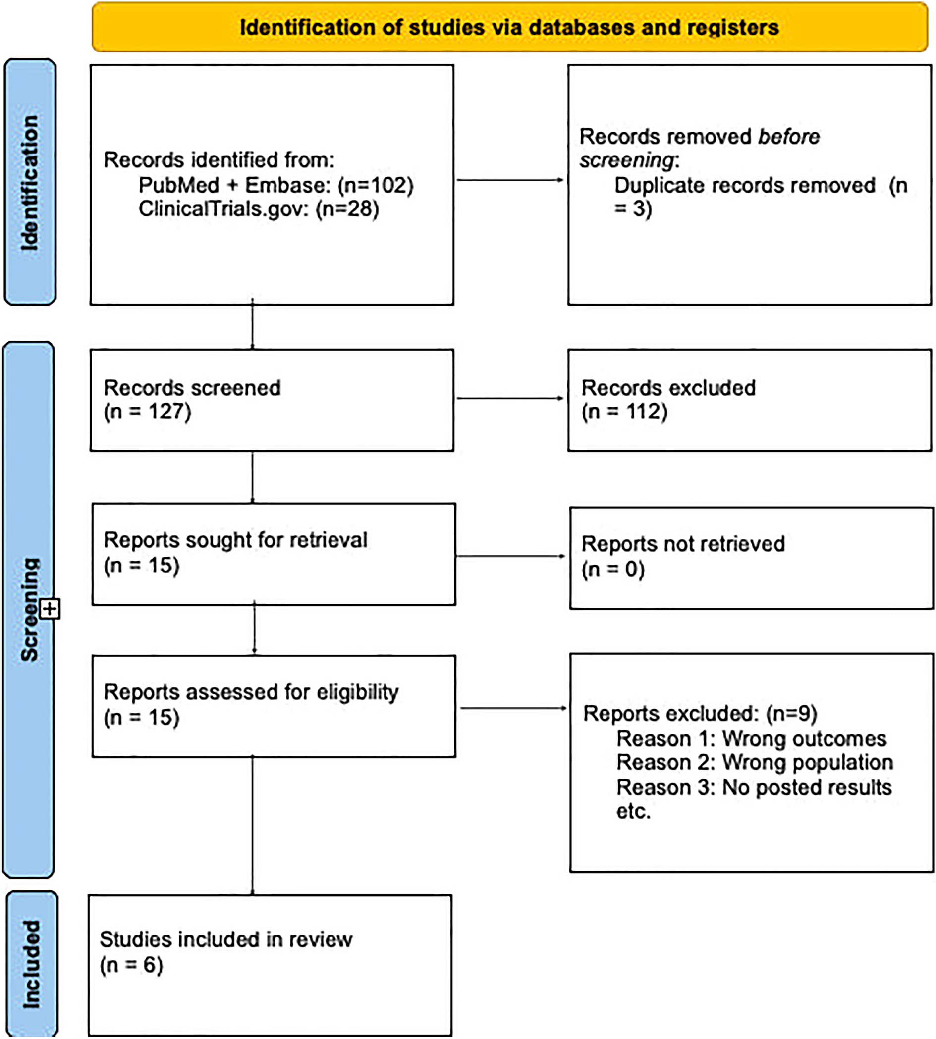
We conducted a retrospective, single-center study at Hôpital Henri Mondor, Assistance Publique–Hôpitaux de Paris (Créteil, France). All adult patients (aged ≥ 18 years) diagnosed with laboratory-confirmed RSV infection between January 2019 and March 2025 were screened. Patients were included if they had a positive RSV polymerase chain reaction (PCR) result from a nasopharyngeal swab and if they had a chest CT scan performed within 7 days of RSV diagnosis or within 72 h prior to diagnosis. If a patient had multiple positive PCR samples, the first positive sample was used for inclusion. Patients with documented co-infection by another respiratory virus or under legal protection were excluded.
Clinical, Biological and Microbiological Data
The following data were recorded: demographic data (i.e., age, sex, weight, height, body mass index, BMI), previous comorbidities (i.e., diabetes, hypertension, chronic respiratory or cardiac disease, chronic renal failure, cirrhosis, immunosuppression), and vaccination status. For patients who are immunocompromised, the type of immunosuppression was specified (i.e., solid organ transplant, long-term corticosteroid use (≥ 20 mg/day prednisone equivalent for ≥ 4 weeks), HIV infection, active solid cancer within the last 5 years, active hematological malignancy within the last 5 years, allogeneic stem cell transplant, immunosuppressive drugs exposure). Microbiological data included bacterial co-infections, while biological data included leukocyte, neutrophil and lymphocyte counts. Data on treatments received during hospitalization (i.e., corticosteroids, ribavirin, palivizumab, intravenous immunoglobulins) and clinical outcomes (i.e., ICU admission, length of stay, severity according to the World Health Organization (WHO) clinical progression scale [17]) were also collected. Clinical factors of severity were recorded, including age ≥ 60 years, chronic heart failure classified as New York Heart Association (NYHA) class III or IV, COPD, chronic respiratory failure requiring long-term oxygen therapy, and/or any preexisting immunosuppression, as previously described [18].
Computed Tomography Assessment
Chest CT scans were acquired using a Revolution CT scanner (General Electric, USA) with a standard protocol involving nonenhanced helicoidal volumetric acquisition from the lung apices to the iliac crest, with automated modulation of the tube current (mAs) and kilovoltage (kV). Pulmonary CT angiography was performed using a dual-energy protocol (80/140 kV) after intravenous injection of iodine contrast media (Iomeron 350, Bracco, France) in case of clinical suspicion of pulmonary embolism. A radiologist (Y.Z.) with 5-year experience in lung imaging reviewed all CT scans independently and was blinded to the clinical data and co-infection status. This review was conducted using a predefined reading grid. The following radiological patterns were assessed systematically: ground-glass opacities, consolidations, bronchiolar disease and interstitial involvement, as well as their distribution and appearance.
Clinical and Radiological Severity Scores
Clinical severity was assessed using the WHO clinical progression scale, which ranges from 0 (no respiratory support) to 10 (death) [17]. According to our study protocol, severe disease was defined as a WHO score of at least 5, which corresponds to the need for supplemental oxygen via a mask or nasal prongs. Radiological severity was quantified using the Chest CT Score (CCTS), which is a semi-quantitative score based on the extent of parenchymal involvement in each of the five lung lobes, initially approved for assessment of severity of coronavirus disease 2019 (COVID-19) CT findings [19]. Each lobe was visually scored from 0 to 5 as follows: 0 (0% involvement), 1 (< 5%), 2 (5–25%), 3 (26–49%), 4 (50–75%), and 5 (> 75%). The sum of the lobe scores yielded a CCTS total ranging from 0 to 25.
Statistical Analysis
Results were expressed as median and 25th and 75th quartiles [Q1–Q3] for quantitative data, and as numbers and percentages for categorical data. Comparisons between groups were made using the Mann–Whitney U test for continuous variables, and either Pearson’s chi-square or Fisher’s exact test for categorical variables. Two-tailed p values < 0.05 were considered statistically significant.
To explore the associations between radiological findings and clinical severity (WHO clinical progression scale ≥ 5) and to avoid collinearity between CCTS and its component radiologic patterns, two multivariable logistic regression models were built (model 1 and model 2). Covariates with p < 0.05 in univariate analysis were included, along with clinically relevant variables meeting a p-value threshold of 0.20. Model 1 included age ≥ 65 years, immunosuppression, and CCTS > 6. In model 2, the CCTS variable was replaced by individual radiological components (i.e., ground glass opacity, consolidation, mucoid impaction, and emphysema). The results are reported as adjusted odds ratios (aOR) with 95% confidence intervals (CI).
Comparative tables were generated using stratifications based on PCR cycle threshold (Ct) values, reflecting viral load, co-infection status, immunosuppression, and CCTS. Spearman correlation coefficients were used to assess correlations between continuous and binary variables. Statistical analyses were performed using R software (version 4.4.2, R Foundation for Statistical Computing, Vienna, Austria).
Ethics Approval
This study received ethical approval from the Ethics Committee of the Société de Réanimation de Langue Française (SRLF): reference CE SRLF 25-019. In accordance with French regulations, this retrospective study, which was based on routinely collected data, was conducted under the legal framework of “non-opposition” to research use. Patients or their representatives were informed through the hospital’s standard procedures for providing information, including the department’s public registry of ongoing research projects, which is available online at https://reamondor.aphp.fr/recherches-sur-donnees-hors-loi-jarde/, and the patient information guide provided upon admission.


Comments (0)