
Remember me
The analysis of water content across treatments revealed significant differences (p ≤ 0.05), indicating the impact of silicon supplementation on the reduction of hyperhydricity. Hyperhydric Growtek-grown shoots had the highest water content (Cv1-HHG, 86.0%), while hyperhydric in vitro sigma-grown shoots had the second highest water content (Cv1-HHS, 77.0%). After 4 wk, the potassium silicon treated shoots on Growtek (liquid medium) Cv1-SiG1 had a water content of 75.0%, while on Sigma (semi-solid medium) Cv1-SiS1 was 70.3%. Both displayed a moderate decrease in water content following silicon treatment. Growtek (Cv1-SiG2) and Sigma (Cv1-SiS2) had a water content of 63.0% and 61.6%, respectively, after 8 wk, which were statistically distinct from all other treatments, highlighting their effectiveness in mitigating hyperhydricity. These results confirmed that silicon, particularly at higher concentrations, significantly reduced excess water accumulation in lingonberry shoots, thus alleviating hyperhydric symptoms (Fig. 1).
Figure 1.
Water content hyperhydric shoots of lingonberry (Vaccinium vitis-idaea L.) genotype Cv1, in liquid (Growtek; Cv1-HHG), and semi-solid (Sigma; Cv1-HHS) and after 4 wk and 8 wk of silicon treated non-hyperhydric shoots in liquid (Growtek; Cv1-SiG1, Cv1-SiG1 respectively), and semi-solid (Sigma; Cv1-SiS1; Cv1-SiS2 respectively). Data represents the mean ± standard deviation (SDs) Experiments were performed in triplicate, and significant differences were assessed using Tukey’s HSD test at P ≤ 0.05. Different letters denote significant differences as determined by Tukey’s HSD test.
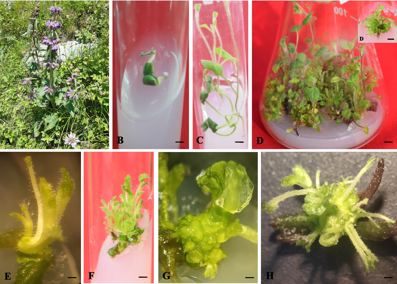
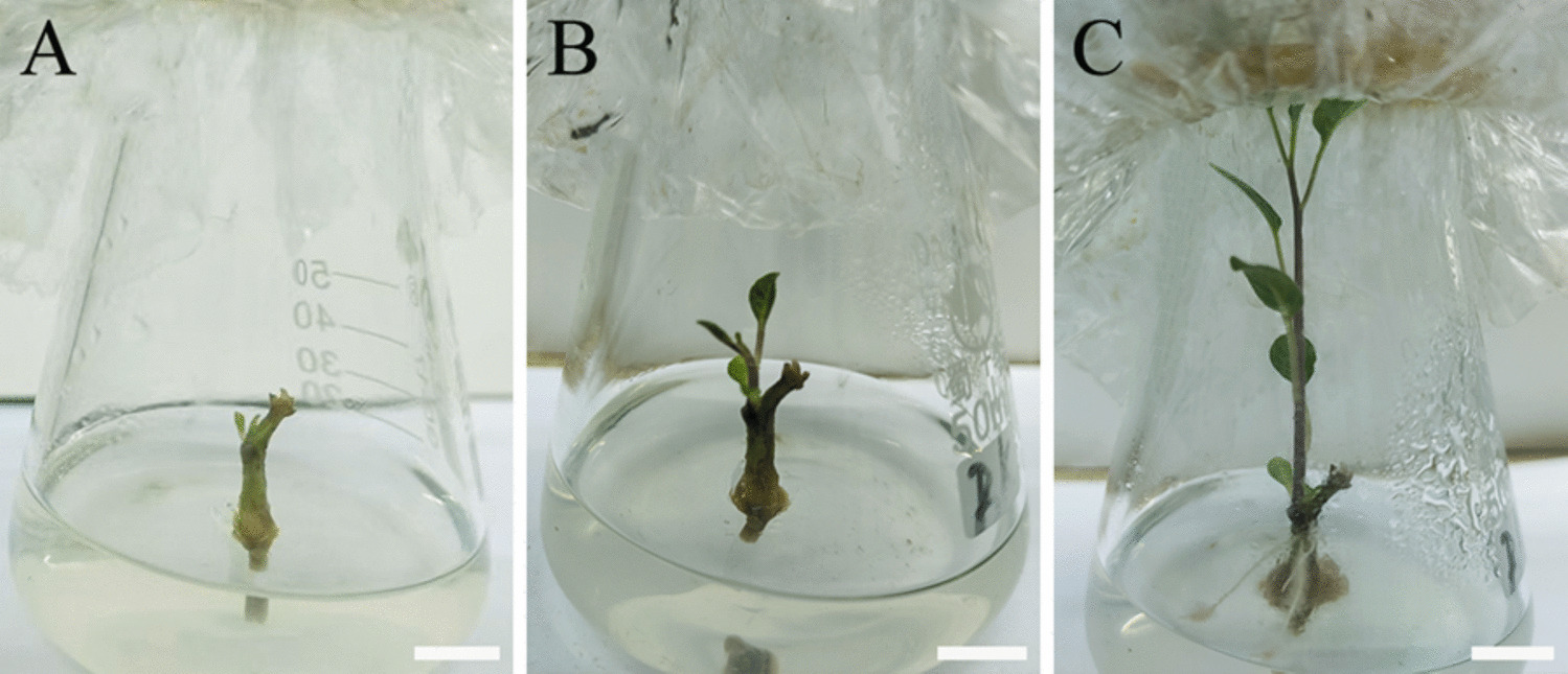
Morphology and hyperhydricity recovery in micropropagated lingonberry shootsHyperhydricity is a significant challenge during micropropagation of lingonberries, with visibly altered plant morphology observed under both liquid (Growtek) and semi-solid (Sigma) media. As seen in Fig. 2A and B, affected shoots exhibited classic hyperhydric symptoms like thickened, blackish, and brittle stems, short internodes, and translucent, curled, and fragile leaves. In severe cases, shoots became twisted, blackened, and necrotic, with a stunted, glassy appearance, and began drying out from the third wk of culture, especially in liquid conditions. In order to mitigate the problem, 1.2 mM K₂SiO₃ was introduced into the medium after 3 wk. While necrotic tissues did not revive, partially affected shoots began showing signs of improvement after 4 wk of silicon treatment (Fig. 2C, D). Gradually, new green shoots emerged from the surviving bases, with a marked improvement in leaf architecture and turgor in both media types. By the 16th wk, a clear and sustained recovery of hyperhydric shoots were observed in both the media (Fig. 2E, F). The silicon treated shoots developed healthy, non-glassy leaves, elongated internodes, and a vigorously growing shoot system, visually distinct from their earlier hyperhydric state. Among all tested concentrations, 1.2 mM K₂SiO₃ emerged as the most effective for reversing hyperhydricity, supporting steady shoot regeneration and physiological balance. This recovery trajectory from initial damage to visible renewal over 16 wk underscored the potential of silicon as a targeted, non-toxic intervention to mitigate culture induced disorders in micropropagated lingonberry.
Figure 2.
Hyperhydric (control) and potassium silicate treated non-hyperhydric shoots of microprapagated lingonberry (Vaccinium vitis-idaea L.) (Cv1). (A) 3-wk-old hyperhydric shoots grown in Growtek (liquid) medium showed darkened, brittle stems and poorly developed shoots with blackish discoloration. (B) in Sigma jar (semi-solid) medium, shoots displayed short internodes with translucent, curled, and fragile leaves. (C) after four wk of potassium silicate treatment, non-hyperhydric shoots in liquid medium began developing new shoots with tiny emerging leaves (D) similarly early recovery were observed in semi-solid medium. (E) healthy, non-glassy leaves, elongated internodes, and a vigorously growing shoot system in liquid and (F) semi-solid medium after 16 wk.
Scanning electron microscopy (SEM)SEM provided detailed insight into the cellular level differences between hyperhydric, and silicon treated micropropagated lingonberry shoots. It revealed distinct anatomical changes associated with physiological disorder and the subsequent recovery.
Leaf surface and epidermal architectureIn hyperhydric shoots cultured in Growtek (liquid) and Sigma (semi-solid) media, SEM analysis of the leaf surface revealed severe anatomical distortions. Epidermal cells were disorganized, with irregular surface textures, loss of cell definition, and distended shapes, consistent with excessive water retention. The stomata were thickened and disoriented in shapes (Fig. 3A, C). Non-glandular trichomes, which play a protective role in normal tissues, were notably fewer in number, deformed, or curled into coiled structures (Fig. 3E, G), making them functionally compromised. These trichomes lacked the structural integrity required for proper defense and transpiration regulation.
Figure 3.

Scanning electron microscopy of lingonberry (Vaccinium vitis-idaea L.) genotype Cv1 showing hyperhydric (control) and silicon treated (non-hyperhydric) tissues. (A) mesophyll tissue with disoriented and thickened stomata in leaf cultured on Growtek medium (liquid) after 3 wk. (B) organized mesophyll tissue and healthy stomata in silicon treated micropropagated shoots on liquid medium after 4 wk. (C) mesophyll tissue with disoriented and thickened stomata in leaf cultured on Sigma jar (semi-solid) medium after 3 wk. (D) organized mesophyll tissue and healthy stomata in silicon treated shoots on semi-solid medium after 4 wk. (E) coiled and disoriented trichomes in liquid medium. (F) well-developed and organized trichomes in silicon treated shoots in liquid medium. (G) coiled and disoriented trichomes in semi-solid medium. (H) well-developed and organized trichomes in silicon treated shoots in semi-solid medium. (I) stem surface of hyperhydric shoots in liquid medium. (J) stem surface of silicon treated healthy shoots in liquid medium. (K) stem surface of hyperhydric shoots in semi-solid medium. (L) stem surface of silicon treated healthy shoots in semi-solid medium. (M) stem cross-section showing irregular xylem and phloem with large intercellular spaces under hyperhydric conditions in liquid medium. (N) normal xylem and phloem without enlarged intercellular spaces in silicon treated shoots in liquid medium. (O) stem cross-section showing irregular xylem and phloem with large intercellular spaces under hyperhydric conditions in semi-solid medium. (P) normal xylem and phloem in silicon treated shoots in semi-solid medium.
By contrast, silicon treated hyperhydric shoots after four wk of 1.2 mM potassium silicate supplementation displayed remarkable anatomical restoration. Newly formed leaves exhibited well-organized epidermal cells, a smoother, well-defined surface, and healthy, erect non-glandular trichomes with normal morphology (Fig. 3F, H). Stomatal development was also notably improved (Fig. 3B, D), with distinct, elliptical stomata and well-formed guard cells, suggesting better regulation of gas exchange. These improvements were consistently observed in both media, highlighting silicon’s role in restoring cellular structure and surface morphology.
Stem anatomy and vascular bundle organizationSimilar patterns were observed in stem cross-sections. In hyperhydric tissues, stems showed disoriented and malformed trichomes, with reduced density and coiled shapes, mirroring leaf abnormalities (Fig. 3I, K). More critically, SEM images revealed disrupted vascular organization: irregularly arranged xylem and phloem (Fig. 3M, O), thin and fragile cell walls, and excessively enlarged intercellular spaces (Fig. 3M, O). These structural deficiencies contributed to the spongy texture of hyperhydric stems and likely impaired water and nutrient transport, reinforcing the visible stunted growth and necrosis seen macroscopically. After silicon treatment, stems exhibited restored trichomes, and surface orientation (Fig. 3J, L), vascular integrity, with well-organized vascular bundles, compact xylem and phloem alignment, and thicker, structurally sound cell walls (Fig. 3N, P). The intercellular spaces were reduced, and trichomes on the stem surface regained their elongated, non-coiled form, suggesting improved tissue rigidity and water conducting efficiency.
Optimization of UHPLCUHPLC-MS analysis was optimized for the detection and quantification of flavonoids, anthocyanins, and phenolic acids using a binary gradient system, coupled with a Triple Quadrupole Mass Spectrometer (TQ-MS), and operated in Dynamic Multiple Reaction Monitoring (dMRM) mode. Chromatographic separation was achieved using a reverse phase column maintained at 30 °C, with a total run time of 12.5 min. A binary solvent system was used comprising water with 0.1% formic acid (Solvent A) and methanol or acetonitrile with 0.1% formic acid (Solvent B), delivered at a constant flow rate of 0.3 mL min−1. The elution gradient started at 80% A and 20% B, transitioned to 15% A and 85% B at 7.3 min, and returned to initial conditions at 8 min, optimizing compound separation and reducing peak broadening and retention time shifts. The injection mode was set to injection with needle wash using a 3 µL injection volume, with a draw speed of 100 µL min−1, ejection speed of 400 µL min−1, and wash time of 3 s. These conditions ensured minimal sample carryover and high reproducibility. The TQ-MS system utilized Agilent Jet Stream Electrospray Ionization (AJS-ESI) in both positive and negative modes. Source parameters were finely tuned for each polarity, and compound specific transitions, fragmentor voltages, and collision energies were optimized to ensure sensitive and selective quantification. A calibration curve with multi-level external calibration standards across twelve concentration levels, ranging from 5 ng mL−1 to 10,000 ng mL−1 was used. Regular maintenance including solvent quality checks and system suitability testing ensured method robustness throughout the study.
Phytochemical profiling of hyperhydric and non-hyperhydric lingonberry shootsUHPLC-MS/MS analysis was conducted to assess the impact of hyperhydricity and silicon based recovery on secondary metabolite accumulation in lingonberry. Ten key phytochemicals, classified into anthocyanins, flavan-3-ols, flavonols, phenolic acids, and procyanidins, were quantified. Statistical analysis using ANOVA followed by Tukey's HSD test revealed significant differences across treatment groups (p ≤ 0.05), with detailed comparisons presented below (Table 2 and Fig. 4). UHPLC-MS/MS chromatograms are presented in supplementary Figure S1.
Table 2. Comparison of hyperhydric and non-hyperhydric (Silicon treated) lingonberry (Vaccinium vitis-idaea L.) plants in both culture media (Growtek: liquid and Sigma: semi-solid)Figure 4.
Phytochemical profiles of micropropagated lingonberry (Vaccinium vitis-idaea L.) shoots under different hyperhydric and silicon treatment conditions. The bar plots represent the concentration (mg kg.−1 FW) of key secondary metabolites categorized as anthocyanins (cyanidin-3-glucoside), flavan-3-ols ((+)-catechin and (−)-epicatechin), flavonols (rutin and quercetin), phenolic acids (chlorogenic acid, gallic acid, vanillic acid), and procyanidins (B1 and B2). Treatments include hyperhydric plants grown in Growtek (liquid) medium (Cv1–HHG), hyperhydric plants in Sigma jar (semi-solid) medium (Cv1–HHS), silicon treated plants in Growtek (Cv1–SiG), and Sigma (Cv1–SiS) medium. Results are expressed as mean ± standard deviation of three biological replicates. Significant reductions in most compounds were observed after silicon treatment compared to hyperhydric tissues (ANOVA, Tukey’s HSD, p ≤ 0.05).
Anthocyanin contentThe concentration of cyanidin-3-glucoside was significantly elevated in hyperhydric tissues, peaking at 21.83 ± 0.13 mg kg−1 FW in Growtek (Cv1-HHG) and 19.46 ± 0.20 in Sigma (Cv1-HHS) hyperhydric shoots. Silicon treatment markedly reduced these levels to 11.66 ± 9.49 (Cv1-SiG) and 10.70 ± 0.03 (Cv1-SiS). All differences were statistically significant (p ≤ 0.05).
ProcyanidinsBoth procyanidin B1 and B2 exhibited strong and significant upregulation in hyperhydric conditions. Procyanidin B1 reached 9.31 ± 8.06 and 8.67 ± 8.09 in Growtek and Sigma hyperhydric shoots, respectively, and was reduced to 6.18 ± 8.36 and 2.53 ± 2.44 following K₂SiO₃ treatment. Procyanidin B2 showed the highest accumulation among procyanidins, with 169.93 ± 27.90 (Cv1-HHG) and 137.08 ± 22.60 (Cv1-HHS), dropping significantly upon Si application to 97.24 ± 15.70 (Cv1-SiG) and 92.73 ± 13.80 (Cv1-SiS), indicating partial biochemical normalization.
FlavonolsAmong flavonols, quercetin content increased significantly in hyperhydric samples (18.22 ± 1.41 in Cv1-HHG and 15.63 ± 1.22 in Cv1-HHS) and was reduced after K₂SiO₃ treatment to 10.53 ± 0.80 and 9.99 ± 0.63 in Growtek and Sigma, respectively. Rutin displayed a similar pattern, with hyperhydric levels at 14.55 ± 1.34 in Cv1-HHG, which significantly declined in the K₂SiO₃-treated group (5.33 ± 0.62) and showed a more dramatic drop in Cv1-HHS (4.07 ± 0.41) and its corresponding Si treatment (3.33 ± 0.19). The decrease was statistically significant (p ≤ 0.05), though more variable in Sigma conditions.
Flavan-3-ols(+)-Catechin exhibited one of the highest responses, increasing more than threefold in hyperhydric conditions (546.86 ± 65.30 in Cv1-HHG and 544.16 ± 49.30 in Cv1-HHS) compared to its reduced values under K₂SiO₃-treatment (183.62 ± 7.8 and 190.62 ± 9.30, respectively). Similarly, (−)-epicatechin was highly elevated in Cv1-HHG (358.69 ± 32.40) and Cv1-HHS (203.38 ± 15.90) and significantly decreased with K₂SiO₃-treatment (198.20 ± 14.60 and 171.27 ± 13.30).
Phenolic acidsChlorogenic acid was the most abundant phenolic compound detected, with concentrations as high as 767.91 ± 63.20 in Cv1-HHG and 522.05 ± 63.00 in Cv1-HHS. K₂SiO₃-treated shoots showed a substantial decline to 256.10 ± 20.60 (Cv1-R1G) and 204.72 ± 19.5 (Cv1-R2S). Vanillic acid also followed this trend, increasing in hyperhydric tissues (2.86 ± 2.44 in Cv1-HHG and 2.13 ± 1.77 in Cv1-HHS) and decreasing with K₂SiO₃-treatment (1.42 ± 1.25 and 1.25 ± 1.05), though with larger standard deviations. In contrast, gallic acid remained stable across all treatments, ranging narrowly from 0.35 ± 0.02 to 0.38 ± 0.03 mg kg−1 FW, and did not show significant differences (p > 0.05). Overall, 9 out of the 10 tested compounds showed statistically significant increases in hyperhydric conditions (p ≤ 0.05), with partial but significant biochemical restoration observed in silicon treated groups. Gallic acid was the only compound that did not exhibit statistically significant variation across treatments. The results suggested a strong link between hyperhydricity and elevated accumulation of antioxidant related phytochemicals, particularly chlorogenic acid, catechins, procyanidins, and flavonols, and indicated the potential of silicon supplementation to modulate these biochemical responses.
Gene expression analysis of hyperhydricity related genes using RT-qPCRIn the present study, RT-qPCR was performed to investigate the expression of 10 genes associated with oxidative stress, ethylene signaling, and metabolism in normal and hyperhydric lingonberry shoots. The analysis used the 2^(-ΔΔCt) method with Actin as the reference gene. Statistical significance was determined using ANOVA followed by Tukey’s HSD (p ≤ 0.05). The gene expression heatmap of 10 genes in both media are shown in Fig. 5.
Figure 5.
Heatmap representation of relative gene expression levels (Ct values) of hyperhydricity related genes in lingonberry (Vaccinium vitis-idaea L.) shoots under hyperhydric and silicon treated conditions in two types of media (liquid and semi-solid). Gene expression was analyzed using RT-qPCR across four groups: silicon treated shoots in Growtek (liquid) (Cv1–SiG) and Sigma jar (semi-solid) (Cv1–SiS) media, and hyperhydric shoots in liquid (Cv1–HHG) and semi-solid (Cv1–HHS) media. The color gradient represents Ct values, where darker blue indicates lower expression and lighter pink or red indicates higher expression. Genes associated with oxidative stress (for example CAT1, SOD, APX), ethylene signaling (ETR1, ACO1, ACS11), and metabolic regulation (COX2, PER12) were significantly upregulated (p ≤ 0.05, ANOVA with Tukey’s HSD). Non-significant differences (n.s) were observed for ADH12 and GMP, suggesting limited or variable response under hyperhydric conditions.
Eight genes showed statistically significant upregulation in hyperhydric shoots (Cv1-HHG) relative to silicon-treated shoots (Cv1-SiG) when cultured in liquid medium. Among the antioxidant related genes, CAT1 showed the highest expression increase with a 35.0-fold upregulation (ΔΔCt = −5.13, log₂FC = 5.13, p = 0.002), followed closely by APX at 32.0-fold (ΔΔCt = −5.00, log₂FC = 5.00, p = 0.001), SOD at 30.0-fold (ΔΔCt = −4.91, log₂FC = 4.91, p = 0.001), and PER12 at 15-fold (ΔΔCt = −3.91, log₂FC = 3.91, p = 0.003). These results pointed to a strong activation of antioxidant defense systems in response to hyperhydric stress. Ethylene signaling and associated metabolic genes were also significantly upregulated: COX2 and ACO1 showed 35.0-fold (ΔΔCt = −5.13, log₂FC = 5.13, p = 0.001) and 28-fold (ΔΔCt = −4.81, log₂FC = 4.81, p = 0.001) increases respectively, while ETR1 and ACS11 were upregulated by 22-fold (ΔΔCt = −4.46, log₂FC = 4.46, p = 0.001) and 18-fold (ΔΔCt = −4.17, log₂FC = 4.17, p = 0.002). Two genes were identified as statistically non-significant expressions: ADH12 had a modest 1.8-fold increase (ΔΔCt = −0.85, log₂FC = 0.85, p = 0.156), and GMP exhibited a 1.7-fold increase (ΔΔCt = −0.77, log₂FC = 0.77, p = 0.089), indicating that these genes were not consistently altered under hyperhydric condition in this medium. In semi-solid medium, the gene expression trends were remarkably consistent with those observed in liquid medium. Again, eight out of ten genes were significantly upregulated in hyperhydric shoots (Cv1-HHS) compared to silicon treated shoots (Cv1-SiS). The antioxidant genes CAT1, APX, and SOD each showed identical maximum expression of 35-fold (ΔΔCt = −5.13, log₂FC = 5.13; p = 0.001 for CAT1 and APX, and p = 0.002 for SOD), while PER12 maintained significant upregulation at 12-fold (ΔΔCt = −3.58, log₂FC = 3.58, p = 0.001). Ethylene related genes also maintained robust expression: COX2 was upregulated by 28-fold (ΔΔCt = −4.81, log₂FC = 4.81, p = 0.001), ACO1 by 25.0-fold (ΔΔCt = −4.64, log₂FC = 4.64, p = 0.001), ETR1 by 21-fold (ΔΔCt = −4.39, log₂FC = 4.39, p = 0.001), and ACS11 by 20-fold (ΔΔCt = −4.32, log₂FC = 4.32, p = 0.003). Genes that were non-significant in liquid medium also showed non-significant expression changes in semi-solid medium as well, ADH12 had a 1.9-fold increase (ΔΔCt = −0.93, log₂FC = 0.93, p = 0.142) and GMP a 1.6-fold increase (ΔΔCt = −0.68, log₂FC = 0.68, p = 0.093). The results obtained showed consistent and significant transcriptional reprogramming in hyperhydric tissues across both liquid and semi-solid media, with 80.0% of genes showing statistically significant upregulation in both conditions. The gene expression patterns, particularly the strong activation of antioxidants and ethylene signaling pathways, suggested a conserved and medium independent molecular response to hyperhydric stress. Genes, such as CAT1, APX, SOD, ACO1, and COX2, emerged as key transcriptional markers of hyperhydricity, while ADH12 and GMP remained stable, indicating their limited role under the tested conditions (Fig. 5).
Comments (0)