
Remember me
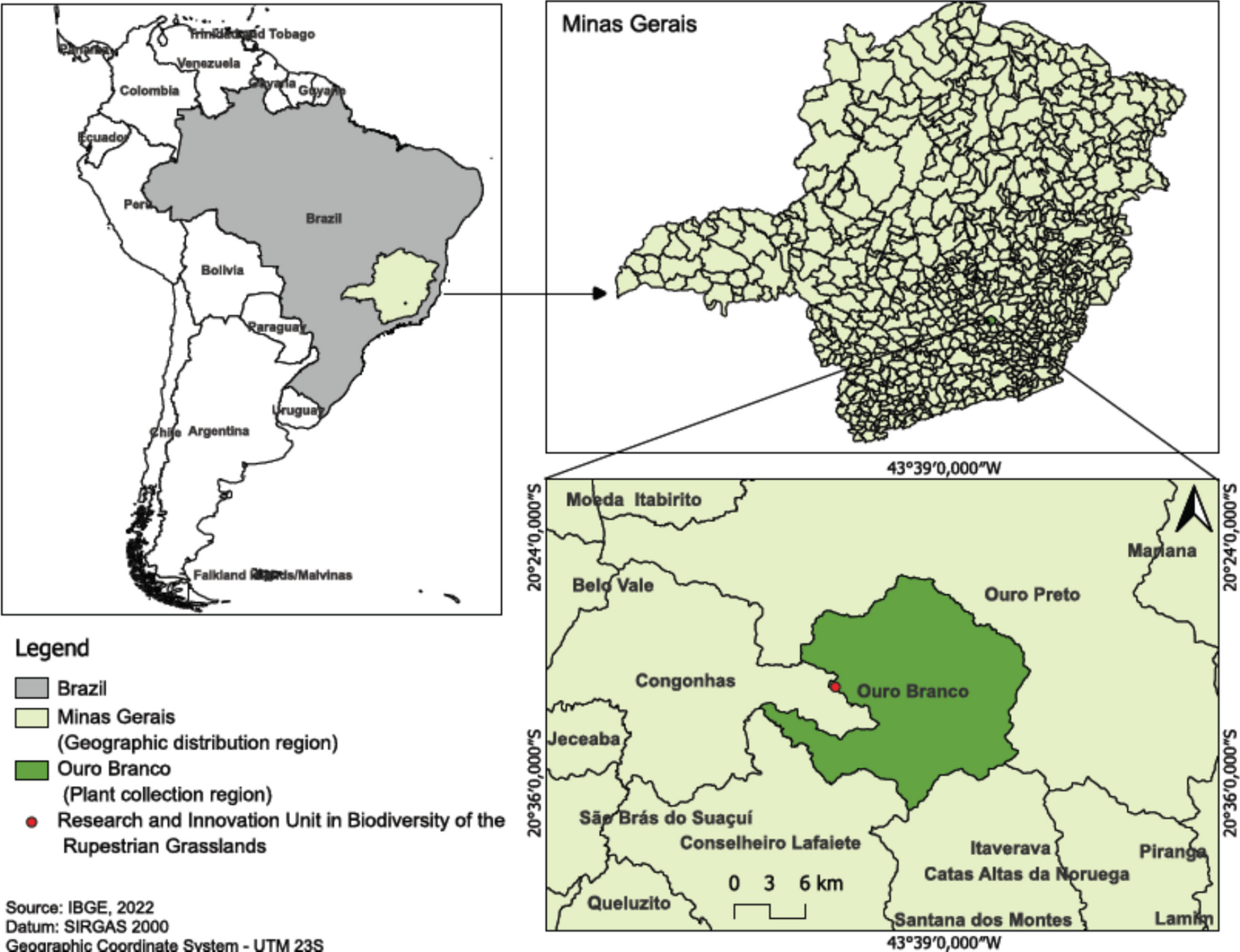
The seeds were collected from wild Sinningia rupicola plants in 2021 (SisGen: ACF88E2), in a Ferruginous Rupestrian Grassland area within the Research and Innovation Unit owned by Gerdau (GERDAU Açominas S.A.), located in the municipality of Ouro Branco, Minas Gerais, Brazil (Fig. 1). Sinningia rupicola seeds were rinsed under running water for 5 min and subsequently immersed in a sodium hypochlorite (NaOCl) solution at 2.0 to 2.5% (v/v) (Clarix®, Viamão, Brazil) for 5, 10, and 15 min inside a horizontal laminar flow hood (VECO, Campinas, Brazil), with the surface previously sterilized with 70% alcohol (Coperalcool®, Barueri, Brazil) and UV light (LUCMAT GL254nm, 30 W, São Paulo, Brazil) for 15 min. After each treatment period, the seeds were rinsed five times with deionized and autoclaved water and, under aseptic conditions, 10 seeds were inoculated per test tube (25 × 150 mm) containing 10.0 mL of Murashige and Skoog (MS; Murashige and Skoog 1962) culture medium supplemented with 30.0 g L−1 sucrose (Synth Ltda, Diadema, Brazil) and 6.0 g L−1 agar (Merck S.A., Rio de Janeiro, Brazil). The pH of the culture medium was adjusted to 5.8 (± 0.05) prior to agar addition, and the medium was autoclaved in autoclave Digitale 20L (BS Equipamentos Ltda, Varginha, Brazil) at 121 °C and 1.0 kgf cm−2 pressure for 20 min.
Figure 1.
Geographic distribution map and collection site area of Sinningia rupicola (Mart.) Wiehler.
The inoculated seeds were cultivated in a growth chamber at 24 °C (± 1 °C), with a 16-h photoperiod (cool white, fluorescent lamps, HO Sylvania T12, 20 W, São Paulo, Brazil) and an irradiance of 40 μmol m−2 s−1 (measured with a LI-COR® LI-250A light meter, Lincoln NE). After 30 d, germination percentage, fungal and bacterial contamination were evaluated. Seedlings that germinated without signs of contamination were considered established and remained in the growth chamber for an additional 30 d before being used in subsequent experiments. The experiment was conducted in a completely randomized design with three treatments and 20 replicates per treatment, with each test tube considered an experimental unit.
Effect of Gas Exchange on In Vitro MultiplicationTo evaluate the effect of gas exchange during the multiplication stage, established shoots measuring 0.5 cm in length were used. Four shoots were subcultured per 250.0 mL glass jar containing 40.0 mL of MS culture medium supplemented with 30.0 g L−1 sucrose (Synth Ltda), 0.5 mg L−1 6-benzylaminopurine (BAP – Sigma®), 0.05 mg L−1 α-naphthaleneacetic acid (NAA – Sigma®), and 6.0 g L−1 agar (Merck S.A., Rio de Janeiro, Brazil). The jars were sealed using three different gas exchange systems: (a) polypropylene caps without membranes (0Mem); (b) polypropylene caps with a single 1.0 cm diameter hole covered with a 1.0 cm2 membrane (1Mem); and (c) polypropylene caps with three 1.0 cm diameter holes, each covered with a 1.0 cm2 membrane (3Mem). The membranes were composed of three layers of microporous surgical tape (Cremer®, Blumenau, Brazil) and one layer of polytetrafluoroethylene (PTFE – Amanco®, Joinville, Brazil) tape measuring 0.05 mm ± 0.01 mm, as described by Saldanha et al. (2012).
After 30 d, the following parameters were evaluated: tissue oxidation, vigor, fungal and bacterial contamination, number of leaves per explant, cluster diameter (bud cluster), and foliar content of photosynthetic pigments [chlorophyll a, chlorophyll b, total chlorophyll (a + b), and carotenoids]. Microbial presence was assessed as either present or absent, while vigor and oxidation were evaluated based on the scales described by Souza et al. (2018). The experiment was conducted in a completely randomized design with 80 replicates (20 jars containing 4 shoots each), with each shoot considered an experimental unit.
Effect of Gas Exchange on In Vitro ElongationClusters measuring 5 mm in diameter were subcultured into 250.0 mL glass jars containing 40.0 mL of MS culture medium supplemented with 30.0 g L−1 sucrose (Synth Ltda), 0.05 mg L−1 BAP (Sigma®), 0.5 mg L−1 NAA (Sigma®), and 6.0 g L−1 agar (Merck S.A). The jars were sealed using the gas exchange systems from the previous experiment (0Mem, 1Mem, and 3Mem).
After 45 d of cultivation, tissue oxidation, vigor, fungal and bacterial contamination, number of leaves per explant, cluster diameter (mm), average shoot length (cm), and foliar content of photosynthetic pigments [chlorophyll a, chlorophyll b, total chlorophyll (a + b), and carotenoids] were evaluated. The experiment was conducted in a completely randomized design with 20 replicates, considering each jar containing 4 clusters as the experimental unit.
Effect of Gas Exchange on In Vitro RootingShoots measuring 1.0 cm in length from the elongation experiment were isolated and inoculated into 250.0 mL jars containing 40.0 mL of MS culture medium supplemented with 30.0 g L−1 sucrose (Synth Ltda), 0.05 mg L−1 BAP (Sigma®), 0.1 mg L−1 NAA (Sigma®, Barueri, Brazil), 0.1 mg L−1 indole-3-butyric acid (IBA – Sigma®), and 6.0 g L−1 agar (Merck S.A.). The jars were sealed using the gas exchange systems from previous experiments (0Mem, 1Mem, and 3Mem). After subculturing and sealing, the jars remained in the growth chamber for 60 d. At the end of the experiment, tissue oxidation, vigor, fungal and bacterial contamination, number of leaves per explant, average shoot length (cm), number of roots, and foliar content of photosynthetic pigments [chlorophyll a, chlorophyll b, total chlorophyll (a + b), and carotenoids] were evaluated. The experiment was conducted in a completely randomized design with 80 replicates (20 jars containing 4 shoots each), considering each shoot as an experimental unit.
AcclimatizationIn vitro–produced Sinningia rupicola seedlings were rinsed under running water to remove the culture medium and subsequently planted in 200.0 mL polypropylene containers (Copobras S.A., Guarulhos, Brazil) with drainage holes at the bottom, filled with 100.0 mL of a substrate composed of decomposed pine bark (CarolinaSoil®, Santa Cruz do Sul, Brazil) and vermiculite (BrasilMinerios S.A., São Luis de Montes Belos, Brazil) in a 2:1 (v/v) ratio, arranged in trays and sealed with plastic film. After planting and adding 5.0 mL of water per seedling, the plants were cultivated in a mini-incubator system (Brondani et al. 2012; Brondani et al. 2018). The seedlings were kept in a growth chamber at 24 °C (±1°C) with a 16-h photoperiod (white LED lamps, SuperLed, 6500k, 9 W, OuroLux®, São Paulo, Brazil) and an irradiance of 37 μmol m−2 s−1 (measured with a USB-650 RED TIDE radiometer, Ocean Optics Inc., Dunedin, FL) and were irrigated every 48 h. After 10 days, the containers were uncovered, and survival and fungal contamination were assessed at 30 d. At the end of the acclimatization period, the seedlings were transferred to a greenhouse covered with shade cloth allowing 50% light transmission and irrigated every other d. After 45 days, seedling survival was evaluated, completing the in vitro seedling production stages (Fig. 2).
Figure 2.
Flowchart of the seedling production stages of Sinningia rupicola (Mart.) Wiehler.
Analysis of Photosynthetic Pigments ContentAt the end of the multiplication, elongation, and rooting experiments, 25.0 mg of fresh leaf tissue from each treatment was sampled. The samples were immersed in 5.0 mL of DMSO solution (Sigma®, Barueri, Brazil) for 48 h in the dark (Lichtenthaler 1987). Absorbance was measured in triplicate using a 10 mm path length quartz cuvette in a Genesys 10UV spectrophotometer (Thermo Scientific, São Paulo, Brazil). Readings were taken at wavelengths of 665, 649, and 480 nm, and the concentrations of chlorophyll a, chlorophyll b, total chlorophyll (a + b), and carotenoids were calculated based on the equations described by Wellburn (1994).
Data AnalysisThe data were analyzed for homoscedasticity and normality using Hartley’s test (P > 0.05) and the Shapiro-Wilk test (P > 0.05), respectively. The results were subjected to analysis of variance (ANOVA, P < 0.05), followed by Tukey’s multiple comparison test (P < 0.05). Data analysis was performed using R software version 4.0.3 (2020), employing the easyanova package (Arnhold 2013).
Comments (0)