
Remember me


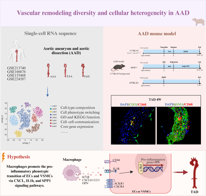
Aortic aneurysm (AA) and aortic dissection (AD) are life-threatening vascular diseases characterized by progressive wall degeneration and structural failure. However, the cellular mechanisms underlying their progression and rupture remain poorly understood. We performed integrative single-cell RNA sequencing analysis on four human aortic datasets, including thoracic aortic dissection (TAD), thoracic aortic aneurysm (TAA), abdominal aortic aneurysm (AAA), and ruptured AAA. We identified disease-specific patterns of immune infiltration and vascular structural cell loss. Vascular smooth muscle cells (VSMCs) and endothelial cells (ECs) were reduced in AA but increased in AD. In ruptured AAA, structural cells were nearly absent, with immune cells predominating. In TAD, VSMCs and ECs exhibited marked phenotypic plasticity, transdifferentiating into multiple cell subtypes. Notably, we identified macrophage-like VSMCs and ECs in AD mouse models. Cell-cell communication analysis revealed that macrophages communicated with VSMCs and ECs via the CXCL, IL1B, and SPP1 signaling axes. Importantly, REL, a member of the NF-κB transcription factor family, was identified as a convergent node integrating these inflammatory signals. This study delineates disease-specific transdifferentiation trajectories of ECs and VSMCs during AA and AD progression. Inflammatory macrophages regulate this phenotypic switching via CXCL, IL1B, and SPP1 signaling, activating REL-driven transcriptional programs that compromise vascular integrity. These findings uncover fundamental differences in cellular heterogeneity and injury mechanisms between AA and AD, offering potential therapeutic targets for preventing disease progression and rupture.
Graphical Abstract The alternative text for this image may have been generated using AI.
Comments (0)