All experiments were performed at 298 K on a Bruker Avance spectrometer (Bruker Instruments, Billerica, MA) operating at 500.03 MHz for 1H and equipped with a 5 mm 1H/BB probe incorporating triple-axis gradient coils. The magnetic field homogeneity on each sample was optimized with an automated 3D field mapping algorithm capable of adjusting up to fifth-order spherical harmonics.
Most chemicals were purchased from Sigma Aldrich (St. Louis, MO, USA) and used without further purification. Vienna Standard Mean Ocean Water 2 (VSMOW2), a widely accepted measurement standard for stable isotope analysis was purchased from the International Atomic Energy Agency (IAEA, Vienna, Austria). All other water samples were obtained from tap water in the greater New Haven area or from bottled water purchased on-line or at local supermarkets. Human plasma samples were obtained during separate DMI studies with intravenous administration of [6,6’-2H2]-glucose or 2H3-acetate, according to previously described infusion protocols [28, 29]. All human studies were approved by the Yale University Institutional Review Board. Plasma samples were obtained prior to and 120 min following the start of substrate administration. Plasma proteins were precipitated with acetone and removed following centrifugation.
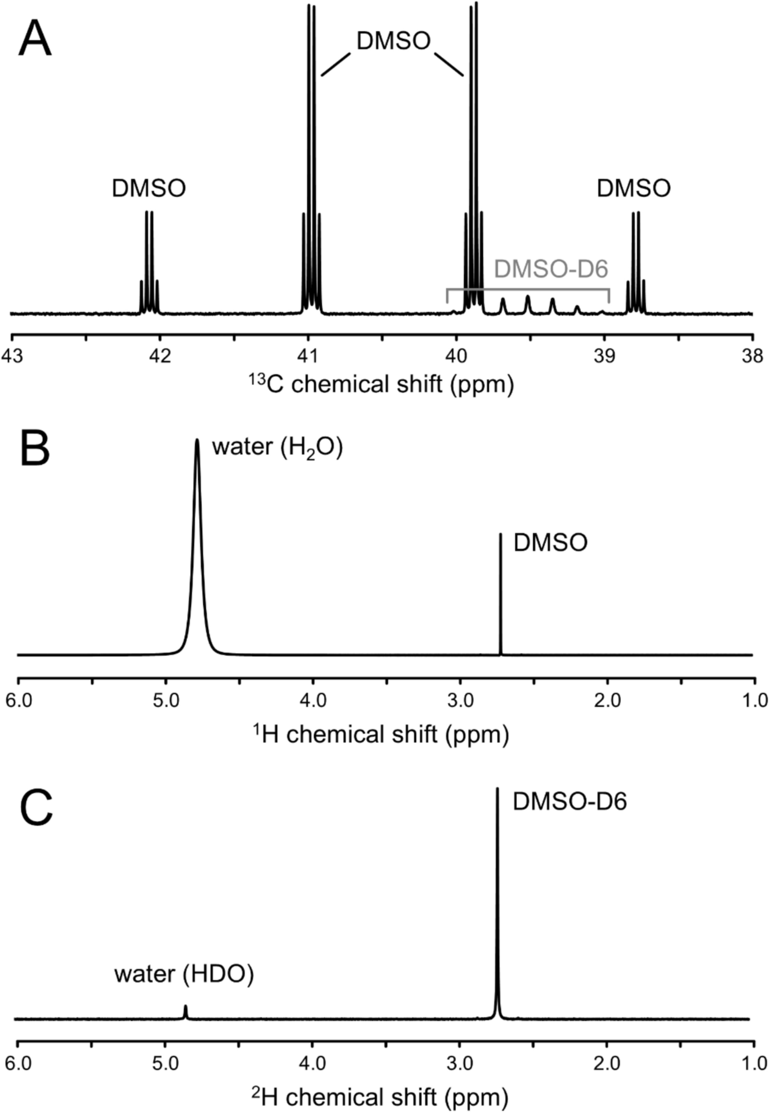
The reference signal was composed of 90% DMSO and 10% DMSO-D6. The exact ratio, Rref, was determined with carbon-13 (13C) NMR, obtained with a 15 µs non-selective (square) excitation pulse followed by 3.2 s acquisition over a 10 kHz bandwidth (32 averages). To minimize spurious signals from decoupling sidebands and eliminate differential signal enhancements all acquisitions were performed without proton decoupling and without nuclear Overhauser enhancement. The long 13C T1 relaxation time constants for deuterated compounds (see Results) mandated a repetition time of 300 s to avoid differential signal saturation. The reference stock solution was kept in the dark at room temperature. Its chemical stability has been verified for up to 3 years through repeated 13C NMR.
Proton (1H) NMR spectra were acquired using a pulse-acquire method (10 µs square pulse excitation) as 8,192 points over a 10 kHz bandwidth with a repetition time of 25 s (2 averages). Most 1H NMR spectra were acquired without a field lock due to the absence of a strong lock signal. Deuterium (2H) NMR spectra were acquired using a pulse-acquire method (250 µs square pulse excitation) as 4,096 points over a 2.5 kHz bandwidth with a repetition time of 5 s (128 averages) using the 2H lock coil and 2H lock amplifier (2 W peak power output). Although 2H NMR spectra were never acquired with a field lock, it was experimentally verified that magnetic field drift did not significantly affect the measurements.
T1 measurements for 2H and 13C NMR were performed with an inversion recovery method using 15–25 delays logarithmically spaced between zero and five times the longest anticipated T1 relaxation time constant. Data were fitted with a single exponential function with three variables (thermal equilibrium magnetization M0, initial magnetization Mz(0) and relaxation time constant, T1). To avoid radiation damping effects, T1 measurements for 1H were based on a saturation recovery method, whereby data was fitted using a two parameter (M0 and T1), single exponential function.
Time-domain signals were zero-filled, Fourier transformed and phase-corrected using in-house developed software written in Matlab (version 9.10 (R2021a), The Mathworks, Natick, MA, USA). Following extensive initial studies, the use of numerical integration and simple spectral line fitting was deemed inadequate in providing robust and consistent signal areas. Reliable signal quantification was achieved through the use of Hankel Singular Value Decomposition (HSVD). Following determination of the center frequencies for water and DMSO, each signal was reconstructed following HSVD using the singular values with frequencies within ± 80 Hz (for 1H) or ± 15 Hz (for 2H) of the center frequencies. These ranges were sufficiently large to include the 13C satellite signals for DMSO. The 2H water enrichment was then calculated from the first datapoint of the reconstructed HSVD FID using Eq. (8). Matlab scripts for HSVD-based quantification, 2H enrichment calculation (i.e., Eqs. (1)-(8)) and example datasets are provided on-line via the Open Science Framework (OSF) repository [30].
When only small quantities of water are available (e.g., plasma from rodent studies), the water can be supplemented with an aprotic solvent to reach the minimum sample volume (i.e., 600 µL for a standard 5 mm diameter NMR tube). Requirements for the aprotic solvent are similar to that of the reference, with the additional requirement that the solvent should not overlap with either water or reference signals. Relative signal frequencies for the water-DMSO system were investigated following dilution with acetone, acetonitrile or dioxane for water contents between 0 and 20% (v/v).
The effects of radiation damping were investigated using a novel MR method whereby the amount of detectable signal, and thus the amount of radiation damping, can be limited through slice selection. Reduction of radiation damping has two requirements, namely (1) a small amount of transverse magnetization throughout the sequence and during acquisition and (2) elimination of all longitudinal magnetization. Fig. S1A shows a pulse sequence that achieved both requirements; the adiabatic excitation pulse ensures that the longitudinal magnetization is reduced to zero. The subsequent magnetic field gradient leads to a reversible phase dispersal across the sample, thereby causing a small, macroscopic transverse magnetization to be maintained throughout the double-spin-echo part of the sequence. The refocusing pulses select a spatial slice along the length of the NMR tube, thereby leading to a permanent reduction of the detectable magnetization.
The method outlined in the Theory section (Eqs. 1,2,3,4,5,6,7) was validated by (1) comparison to the known composition provided by the VSMOW2 standard and (2) comparison with results obtained using a double-compartment NMR tube, consisting of a 4 mm diameter NMR tube inserted into a standard 5 mm diameter NMR tube. The outer compartment functions as a reference signal to account for coil loading differences. The composition can be flexible provided that the compartment contains a 2H-labeled compound that does not overlap with the signal from deuterated water. Here the outer compartment contained DMSO/DMSO-D6 (90/10% (v/v)). The inner compartment is used for two separate experiments containing (1) ‘pure 100%’ deuterium oxide (D2O) or (2) the sample with a to-be-determined 2H composition. The ratio of 2H water signal, Swater,exp2/Swater,exp1, scaled for differences in coil loading, is equal to the 2H enrichment of the sample. Assumptions for the double-tube approach are identical B1 distributions for the two experiments and 100% 2H enrichment for the first experiment. Note that the double-compartment NMR tube method is also independent of the amount of reference. However, unlike the proposed method, the double-compartment NMR tube method requires specialized hardware (i.e., non-standard NMR tubes) and is not practical for large number of samples measured in an automated fashion.
All statistical comparisons used a two-sided Wilcoxon signed rank test whereby p < 0.05 was considered to indicate a statistically significant difference. Statistical analysis was performed using the statistical toolbox in MATLAB (version R2021a; MathWorks).


Comments (0)