
Remember me

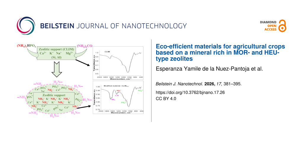

This proof-of-concept study evaluated whether semi-solid extrusion (SSE) 3D printing could be used to fabricate multilayer topical films that simultaneously enhance skin bioadhesion and photoprotection of curcumin, a highly photolabile anti-inflammatory and antioxidant compound. The development of topical films for cutaneous delivery faces several challenges, including the need for strong skin adhesion and the protection of photolabile actives from light exposure. We hypothesized that multilayered films designed for the cutaneous delivery of curcumin and produced by SSE could address these limitations. To overcome its poor solubility and enhance bioadhesion, curcumin was encapsulated in polymeric nanocapsules (C-NCs), yielding a mean particle size of 218 ± 5 nm, a polydispersity index of 0.10 ± 0.02, a zeta potential of −11 ± 4 mV, and 100% encapsulation efficiency. Films were fabricated containing either C-NCs (FC-NC) or unloaded curcumin (FC) and consisted of three layers, namely, a chitosan-based bottom layer, a middle layer of carboxymethylcellulose and alginate, and a carboxymethylcellulose top layer incorporating titanium dioxide (TiO2). The lower and intermediate layers contained C-NC or curcumin. The final films (15 × 15 × 1.5 mm) contained 282.20 ± 7.75 µg and 246.80 ± 6.70 µg of curcumin in FC-NC and FC, respectively. Films containing the bottom chitosan layer exhibited the highest bioadhesion, while the presence of a TiO2 top layer effectively prevented UVC-induced photodegradation, supporting our hypothesis. Furthermore, the presence of C-NCs in FC-NC films promoted higher bioadhesion. This proof-of-concept study demonstrates the feasibility of integrating nanocarriers with 3D printing technology to engineer multilayer polymeric films for cutaneous application, offering enhanced bioadhesion and photoprotection. This work demonstrates how additive manufacturing can be used to design hierarchically structured, nanocarrier-integrated systems with spatially resolved functionalities.
Introduction3D printing is an innovative technology that is poised to transform personalised medicine and healthcare manufacturing. For the healthcare industry, the use of 3D printing provides several key advantages, including the ability to adapt dosages according to a patient’s physiological characteristics and genetic background, as well as their individual response to drugs and active substances. This level of personalisation significantly minimises the likelihood of adverse drug reactions and enhances therapeutic efficacy. Furthermore, a wide range of pharmaceutical formulations, drug combinations, and release profiles can be developed in the form of either immediate-release or extended-release options . Several 3D printing techniques can be applied to pharmaceutical development, including material jetting, fused deposition modelling, stereolithography, selective laser sintering, binder jetting, semi-solid extrusion (SSE), and direct powder extrusion .
SSE is a method in which a semi-solid material is placed into a syringe and continuously extruded layer by layer onto a smooth surface until the entire object is created. The semi-solid material itself consists of polymers, solvents, and excipients blended at an optimal ratio; the mixture should not be overly viscous to enable efficient extrusion from the syringe tip, but it must also not be too fluid in order to support the weight of each subsequent layer. After extrusion, a post-printing drying phase is required to eliminate the solvent. A significant advantage of this technique is that it can be performed at room temperature or even at lower temperatures, making it ideal for thermosensitive materials .
The SSE approach can be applied to produce various pharmaceutical dosage forms and can be tailored to the requirements of specific applications, such as the production of films intended to promote skin healing . Although topical films can be an effective option for facilitating skin repair and wound healing, they must be carefully manufactured to maintain optimal moisture levels, which may vary depending on the wound type, such as venous ulcers or burns . An ideal dressing should exhibit suitable bioadhesive properties, prevent bacterial growth, minimise pain, and control odour, while also being cost-effective and easy to replace. Current research therefore focuses on the development of specialised dressings using materials such as alginate and carboxymethylcellulose as alternatives to traditional gauze .
Bioactive molecules such as curcumin have shown promise for use in the promotion of skin repair and wound healing. Curcumin is a photosensitive polyphenol derived from Curcuma longa (turmeric) with well-documented biological activities , including properties that may be advantageous for skin-healing applications. For example, curcumin exhibits strong anti-inflammatory activity through inhibition of nuclear factor kappa B (NF-κB) signalling, as well as antioxidant activity through reactions with free radicals or reactive oxygen species (ROS) and inhibition of lipid peroxidation and DNA damage. These mechanisms contribute to reduced oxidative stress and enhanced wound-healing processes . Consequently, curcumin is currently under investigation for incorporation into topical dosage forms such as emulsions, fibres, films, and hydrogels for wound-healing applications.
However, curcumin also exhibits low aqueous solubility, which limits its use in certain formulations . Nanoparticles have the potential to overcome this limitation, as they enable the development of formulations with reduced particle sizes (typically between 100 and 500 nm) and tailored physicochemical properties . In the pharmaceutical context, nanoparticles, including polymeric nanoparticles, have been extensively investigated as drug carriers. The use of nanosystems to enhance the bioactivity of curcumin was reported by Krausz et al., who demonstrated that a curcumin-loaded nanosystem inhibited the growth of methicillin-resistant Staphylococcus aureus (MRSA) and Pseudomonas aeruginosa in vitro in a dose-dependent manner, while also improving wound healing and inhibiting MRSA growth in vivo in a murine wound model . More recently, Terzopoulou et al. developed collagen patches containing chitosan nanoparticles loaded with curcumin, which showed efficacy against psoriasis in in vitro assays . Similarly, Olmos-Juste et al. employed 3D printing to produce alginate–cellulose nanofibre patches exhibiting adequate disintegration and in vitro release properties for the local administration of curcumin .
Although curcumin-loaded chitosan or alginate films have been previously reported, no studies have explored the deliberate spatial distribution of functional components enabled by multilayer 3D printing, nor the incorporation of a photoprotective top layer to shield curcumin from UV-induced degradation. Thus, the innovation of the present work lies not in the individual materials themselves, but in their integration into a rationally designed, layer-by-layer system produced through additive manufacturing.
At present, specific strategies to protect curcumin in topical films from UV-induced degradation have not been extensively investigated, and studies focusing on improving the skin-adhesion properties of curcumin-containing topical films remain limited. Therefore, in the present study, we designed 3D-printed topical films comprising three distinct layers, namely, (i) a bottom layer composed of a chitosan gel containing curcumin, (ii) an intermediate layer composed of carboxymethylcellulose, alginate gel, and curcumin, and (iii) a top layer composed of carboxymethylcellulose and titanium dioxide (TiO2). Curcumin was incorporated into the formulations either in its encapsulated form, loaded into polymeric nanocapsules, or non-encapsulated and dissolved in a hydroalcoholic solution. The preparation of films containing curcumin-loaded polymeric nanocapsules or non-encapsulated curcumin was based on previous studies demonstrating the ability of nanocarriers to improve the aqueous solubility and antioxidant activity of curcumin when formulated as a gel .
Consequently, the objective of the present study was to use SSE 3D printing to design and produce, for the first time, three-layer films composed of chitosan, carboxymethylcellulose, and alginate, containing either nanoencapsulated curcumin loaded into polymeric nanocapsules or non-encapsulated curcumin added from solution. The advantages of producing 3D-printed polymeric films compared with previously developed hydrogels for skin delivery include improved control over the applied curcumin dose, as well as the personalisation of the size and shape of the printed films for the treatment of specific skin areas, such as wounds.
In this proof-of-concept study, we hypothesised that SSE 3D printing could be strategically employed to construct multilayer topical films capable of addressing two key limitations of curcumin for cutaneous delivery, namely, (i) its pronounced photosensitivity, which necessitates a protective barrier and (ii) its limited affinity for the skin surface, which requires enhanced bioadhesion to maintain residence time. To test this hypothesis, we designed a trilayer architecture in which a chitosan-rich lower layer provides muco- and bioadhesive properties, while a TiO2-containing upper layer acts as a physical photoprotective barrier, and curcumin is incorporated either in a nanoencapsulated form or as a solubilised active. The scope of the present work did not include in vitro release or permeation studies, as the primary aim was to validate the structural and functional feasibility of a multilayer 3D-printed design; biopharmaceutical evaluations will be addressed in subsequent studies.
Experimental MaterialsCurcumin (purity level: 65%), poly(ε-caprolactone) (PCL), sorbitan monooleate, sodium alginate (≈200–300 kDa), and low-molecular-weight chitosan (50–190 kDa) were purchased from Sigma-Aldrich (São Paulo, Brazil). Sodium carboxymethylcellulose 4,000 cP (Na-CMC) was obtained from Levviale (São Paulo, Brazil). Titanium dioxide in the rutile form (Eusolex® T-AVO) was obtained from Alpha Química. Acetone was obtained from Neon Comercial Ltda (São Paulo, Brazil), grape seed oil was purchased from Delaware Importadora Química (Porto Alegre, Brazil), and polysorbate 80 was purchased from Henrifarma (São Paulo, Brazil). HPLC-grade solvents, acetonitrile and methanol, were acquired from Merck Millipore (Porto Alegre, Brazil). Trifluoroacetic acid was obtained from Sigma-Aldrich (São Paulo, Brazil). All reagents and solvents were of pharmaceutical or HPLC grade and were used as received.
Nanocapsule suspensions PreparationNanocapsules were prepared using a preformed polymer interfacial deposition method that was previously described by our group, but with some modifications . Curcumin-loaded polymeric nanocapsules (C-NC) were prepared using an organic phase composed of 0.010 g of curcumin, 0.1 g of PCL, 27 mL of acetone, 0.0390 g of sorbitan monooleate and 165 µL of grape seed oil, under magnetic stirring at approximately 400 rpm for 4 h at 40 °C. After complete solubilisation of all components, the organic phase was combined by pouring it into the aqueous phase under moderate stirring (the temperature and agitation speed parameters were the same as those of the organic phase). The aqueous phase was composed of 0.077 g of polysorbate 80 and 54 mL of ultrapure water. Acetone was removed under reduced pressure, and the resulting suspensions were concentrated to 10 mL. The experiment was carried out in a dark environment, protected from light. Blank formulations (B-NC) were prepared under the same conditions, without addition of curcumin in the organic phases. All formulations were prepared and characterised in triplicate.
Particle size, pH and zeta potentialThe z-average particle size and the polydispersity index were measured by dynamic light scattering using a Zetasizer® Nano ZS (ZEN 3600, Malvern Instruments, USA). For this analysis, 10 µL of the C-NC formulation was diluted in 5 mL of ultrapure water (previously filtered through a 0.45 µm hydrophilic membrane, Millipore®), corresponding to a 500-fold dilution, as shown in Equation 1. The zeta potential was determined by measuring electrophoretic mobility at 25 °C using the same instrument (Zetasizer® Nano ZS, ZEN 3600, Malvern Instruments, USA). Prior to analysis, the samples were diluted 500-fold in an aqueous 10 mmol/L NaCl solution and filtered through a 0.45 µm membrane (Millipore®). The pH of the formulations was measured without prior dilution at 25 °C using a calibrated potentiometer (VB-10, Denver Instrument, USA).
![[2190-4286-17-30-i1]](https://www.beilstein-journals.org/bjnano/content/inline/2190-4286-17-30-i1.svg?max-width=590&scale=1.18182) (1)
(1)where C2 is the concentration of the sample after dilution, C1 is the initial concentration of the nanocapsule suspension, V1 is the volume of the nanocapsule suspension that was used, and V2 is the final volume of the sample.
Curcumin content and encapsulation efficiencyThe curcumin content was assayed by high-performance liquid chromatography with ultraviolet detection (HPLC-UV) . The chromatographic system consisted of a Shimadzu LC-20A chromatograph (Tokyo, Japan), using a C18 reversed-phase column (250 × 4.60 mm, 5 µm, 100 Å; Zorbax Eclipse Plus, Agilent). The mobile phase was composed of acetonitrile and trifluoroacetic acid (0.1%) (60:40, v/v), and the pH was adjusted to 3.0 using triethanolamine. Chromatography was conducted at a flow rate of 1.0 mL/min, with an injection volume of 20 µL, and curcumin was detected at a wavelength of 427 nm. The method was linear over the concentration range of 2.5–50.0 µg/mL of curcumin (y = 126634x − 12894; r = 0.9994), with intra-day variability lower than 2% and limits of detection and quantification of 0.2 and 0.7 µg/mL, respectively. Method specificity was confirmed by comparing chromatograms of the samples with those obtained from the placebo formulation. The curcumin content was determined after extraction with acetonitrile for 20 min, followed by filtration through a 0.45 µm membrane (Millipore®) prior to HPLC analysis. All analytical procedures were performed under dark conditions to protect the samples from light.
Encapsulation efficiency was determined using an ultrafiltration–centrifugation technique (Amicon® 10,000 MW cutoff, Millipore®, Billerica, USA). Non-encapsulated curcumin was separated from curcumin-loaded nanocapsules using an ultrafiltration device, followed by centrifugation at 2150g for 10 min. Encapsulation efficiency was calculated as the difference between the total curcumin content and the non-encapsulated curcumin content.
Production of the printing InksThree different polymeric hydrogels or hydrogel blends were produced, corresponding to the three distinct layers of the film. Table 1 presents the composition of the different hydrogels prepared and used as printing inks for the present study. Some hydrogels were prepared solely for comparative purposes and were used during the characterisation step. Chitosan was the polymer used to produce the hydrogel for the bottom layer of the film. This natural polymer was initially mixed with glycerine in a porcelain mortar, and either C-NC (curcumin-loaded polymeric nanocapsules) or C (curcumin) solubilised in a hydroalcoholic solution (1:1, v/v) was slowly added. After complete homogenisation, 3% (v/v) acetic acid was added to solubilise the chitosan. For the intermediate layer, hydrogels of sodium carboxymethylcellulose (Na-CMC) and sodium alginate were produced separately. Na-CMC was dispersed in either C-NC or C until complete gelation was achieved. The sodium alginate hydrogel was produced using the same procedure, but with the addition of calcium carbonate to induce gelation. The hydrogels were then combined and mixed in a porcelain mortar at a ratio of 3.5:1 (w/w) Na-CMC/alginate, with the addition of glycerine, and the resulting blend was used as the printing ink. For the top layer, Na-CMC was mixed with glycerine and solubilised in purified water in a porcelain mortar until gel formation was achieved. Titanium dioxide (TiO2) was then added, and the gel mixture was completely homogenised using a pestle.
Table 1: Composition of the printing inks (hydrogels) for the SSE printing process.a
Alginate (%) Na-CMC (%) Chitosan (%) TiO2 (%) Glycerine (%) C-NC (%) C (%) H2O (%) HG-ChiC-NC — — 7 — 10 83 — — HG-CMC/AlgC-NC 5 6 — — 10 79 — — HG-ChiC — — 7 — 10 — 83 — HG-CMC/AlgC 5 6 — — 10 — 79 — HG-Chi — — 7 — 10 — — 83 HG-CMC/Alg 5 6 — — 10 — — 79 HG-CMC/TiO2 — 6 — 1 10 — — 83aHG-ChiC-NC: chitosan hydrogel containing curcumin-loaded polymeric nanocapsules; HG-CMC/AlgC-NC: sodium carboxymethylcellulose and alginate hydrogel blend containing curcumin-loaded polymeric nanocapsules; HG-ChiC: chitosan hydrogel containing curcumin added as hydroalcoholic solution; HG-CMC/AlgC: sodium carboxymethylcellulose and alginate hydrogel blend containing curcumin added as hydroalcoholic solution; HG-Chi: chitosan hydrogel; HG-CMC/Alg: sodium carboxymethylcellulose and alginate hydrogel blend; HG-CMC/TiO2: sodium carboxymethylcellulose and alginate hydrogel blend containing titanium dioxide.
pH measurementsAfter dispersion of each hydrogel in water (10% w/v), the pH of the resulting printing inks was measured at 25 °C using a calibrated potentiometer (VB-10, Denver Instrument, USA).
Rheological analysesRheological properties of all hydrogels were evaluated using an ARES G2 rheometer (TA Instruments, New Castle, DE, USA) equipped with a parallel-plate geometry (25 mm diameter, 0.9 mm gap) at 20 °C. Viscoelastic properties, including the storage modulus (G′), loss modulus (G″), and loss tangent (tan δ), were measured using an oscillatory frequency sweep over an angular frequency range of 1–600 rad/s at a constant strain of 0.5%. Flow behaviour was assessed using a shear rate ramp from 0.1 to 1.0 s−1 and subsequently from 1.0 to 0.1 s−1, in order to determine the best-fit flow model for the hydrogels. An oscillatory amplitude sweep was performed over a strain range of 0.1–500% at a constant frequency of 1 Hz to determine the linear viscoelastic region and to estimate the yield stress. Thixotropic behaviour was evaluated using a three-interval thixotropy test (3ITT), conducted as follows: the first interval was performed at 1.0% strain for 100 s, the second interval at 100% strain for 100 s, and the third interval at 1.0% strain for 100 s. All measurements were performed in triplicate.
Topical skin films 3D printingThree-layer films containing curcumin-loaded polymeric nanocapsules (FC-NC) or curcumin solubilised in a hydroalcoholic solution (FC) were 3D-printed by semi-solid extrusion using a BioEdTech printer (São Paulo, Brazil) and three different hydrogels as printing inks. HG-Chi was used as the printing ink to produce the bottom layer of the film. Subsequently, two intermediate layers were printed using HG-CMC/Alg on top of the bottom layer, followed by the deposition of two upper layers using HG-CMC/TiO2. Because the 3D printer was equipped with a single printing head, the syringe containing the printing ink had to be manually replaced between layers with the syringe containing the hydrogel corresponding to the layer to be printed; this procedure required less than 45 s. The film geometry was designed using PrusaSlicer software (Prusa Research, Prague, Czech Republic), with final dimensions of 15 mm (length) × 15 mm (width) × 1.5 mm (height). The printer recognised the generated model and, using a syringe fitted with a nozzle with a diameter of 0.41 mm, deposited the material layer by layer at room temperature. The printing parameters were set to a rectilinear infill pattern with 100% infill density and an extrusion speed of 6 mm/s. After printing, the films were dried at 25 °C for 48 h to allow for water evaporation.
Physical characterisationThe physical properties of the resulting 3D-printed films (n = 10) were measured. Namely, films were individually weighed on an analytical balance (Shimadzu, Tokyo, Japan), and their length, width and height were measured using a digital calliper (Digimess, São Paulo, Brazil).
Curcumin contentThe curcumin content of the films (n = 3) was determined by transferring a single weighed unit to 10 mL of ultrapure water in a volumetric flask of 25 mL, followed by mixing under magnetic stirring for 1 h, until complete disintegration. After this period, the volume of the volumetric flask was adjusted to 25 mL with methanol and sonicated for 2 h. Then, a 0.5 mL aliquot was removed and transferred to a 5 mL volumetric flask and diluted with the mobile phase. These samples were filtered through a 0.45 µm membrane, and the level of curcumin was assayed by HPLC using the method described in section “Curcumin content and encapsulation efficiency“. The entire assay was conducted under protection from light.
Differential scanning calorimetryDifferential scanning calorimetry (DSC) was performed using a differential scanning calorimeter (Shimadzu DSC-60), at a heating rate of 10 °C/min, from room temperature to 300 °C. N2 gas was used at a flow rate of 50 mL/min. All film components (chitosan, CMC and alginate), as well as the resulting films (FC-NC and FC) were analysed separately.
Skin adhesion measurementsBioadhesion tests of the complete 3D-printed films (FC-NC and FC, n = 3) were conducted using a texture analyser (TA.XT plus, Stable Micro Systems, Godalming, Waverley, UK). To evaluate the influence of chitosan, the bioadhesion of films containing curcumin or nanoencapsulated curcumin without a chitosan layer (FC-NC w/o CH and FC w/o CH, n = 3) was also evaluated using porcine ear skin as model. The ears were donated by Ouro do Sul Cooperative, Harmonia, Rio Grande do Sul, Brazil. The ear skins were cut, removing the hair and adipose tissue. The skins were stored in aluminium foil at −20 °C until the day of the experiment, without using any media or further treatment. On the day of the experiments, skin tissue was maintained under room conditions for at least 30 min prior to the experiments. A volume (20 µL) of ultrapure water was added by pipette onto the tissue to ensure hydration, and excess water was removed with absorbent paper. Skin samples were fixed to the equipment probe with instantaneous adhesive, while films were fixed to the equipment’s platform with double-sided tape. The equipment promoted contact between the skin sample and the film, with a force of 290 mN for 3 min. The probe holding the skin sample was then withdrawn from the surface of the film by the platform at a constant speed of 0.10 mm/s until total displacement was achieved. The work required to detach the skin sample from the 3D-printed films was calculated based on the peak force and maximum displacement after complete detachment using software (Exponent, Stable Micro Systems, Godalming, Waverley, UK) .
Photodegradation behaviourPhotodegradation analyses were conducted in a mirrored chamber under an UVC lamp for 24 h, and all analyses were done in triplicate. UVC irradiation was selected as a stress condition to perform a forced degradation photostability challenge, in line with accelerated conditions commonly employed to quantify intrinsic photolability of highly photosensitive compounds such as curcumin. This approach provides an exaggerated but controlled environment to compare the relative protective effects of the TiO2-containing layer. It was not intended to mimic physiological solar exposure, and future assessments under UVA/UVB spectra are planned. Four groups (a–d) were evaluated as follows: (a) complete films containing nanoencapsulated curcumin (FC-NC), (b) complete films containing unloaded curcumin (FC), (c) films containing nanoencapsulated curcumin (FC-NC) without an upper TiO2-containing carboxymethylcellulose layer, and (d) films containing unloaded curcumin (FC) without an upper TiO2-containing carboxymethylcellulose layer. Films (n = 27) from each group were placed in the UVC chamber for 24 h. At 2, 4, 6, 8, 10, 18, and 24 h, three films were removed from the chamber and underwent the curcumin extraction process and HPLC assay using the methods described in section “Curcumin content and encapsulation efficiency“.
Statistical analysesStatistical analyses were performed using one-way analysis of variance (ANOVA) followed by a post hoc Tukey’s test for multiple comparisons of means. Differences were considered significant at p ≤ 0.05.
Results Nanocapsule suspensionsCurcumin-loaded polymeric nanocapsules (C-NC) and blank nanocapsules without curcumin (B-NC) were produced (n = 3). Table 2 provides a summary of results from physicochemical characterization.
Table 2: Physicochemical properties of curcumin-loaded nanocapsules (C-NC) and corresponding blank nanocapsule (B-NC) formulations.
C-NC B-NC z-average (nm) 218 ± 5 230 ± 8 polydispersity index 0.11 ± 0.02 0.10 ± 0.02 zeta potential (mV) −11 ± 4 −10 ± 1 pH 6.4 ± 0.1 6.9 ± 0.1 encapsulation efficiency (%) 100 ± 0.00 — curcumin content (mg/mL) 0.98 ± 0.03 — Printing inkDuring the 3D printing process, the rheological behaviour of a printing ink is directly related to printability and the ability to form stable, structured layers, in order to avoid collapse after addition of subsequent layers. Because of this, the rheological properties of the hydrogels tested in the present study were investigated, and results are shown in Table 3. Based on a frequency oscillation test, as shown in Figure 1A–C, all samples had G′ values greater than G″ and, hence, a tan δ (ratio of G″/G′) below 1. Flow curves presented as shear stress (τ) and apparent viscosity (η) versus shear rate are shown in Figure 1D–F. Finally, Table 4 shows the thixotropic properties of the hydrogels, demonstrating recovery of the viscosity of the hydrogels after stress cessation.
Table 3: Yield stress presented as the crossover modulus and complex viscosity of the hydrogels at 1 Hz (frequency sweep test). Values are presented as the mean ± SD (n = 3).a
Yield stress (Pa) Complex viscosity (Pa·s) HG-ChiC-NC 332.33 ± 18.18 136.19 ± 12.88 HG-CMC/AlgC-NC 391.34 ± 39.23 193.27 ± 18.77 HG-ChiC 539.89 ± 31.12 310.60 ± 22.07 HG-CMC/AlgC 758.43 ± 180.82 469.51 ± 73.63 HG-Chi 352.45 ± 27.94 102.26 ± 11.69 HG-CMC/Alg 366.90 ± 6.30 187.62 ± 1.53 HG-CMC/TiO2 216.26 ± 12.78 101.55 ± 8.97aHG-ChiC-NC: chitosan hydrogel containing curcumin-loaded polymeric nanocapsules; HG-CMC/AlgC-NC: a blend of sodium carboxymethylcellulose and alginate hydrogel containing curcumin-loaded polymeric nanocapsules; HG-ChiC: chitosan hydrogel containing curcumin in a hydroalcoholic solution; HG-CMC/AlgC: a blend of sodium carboxymethylcellulose and alginate hydrogel containing curcumin in a hydroalcoholic solution; HG-Chi: chitosan hydrogel; HG-CMC/Alg: a blend of sodium carboxymethylcellulose and alginate hydrogel; HG-CMC/TiO2: a blend of sodium carboxymethylcellulose and alginate hydrogel containing titanium dioxide.
![[2190-4286-17-30-1]](https://www.beilstein-journals.org/bjnano/content/figures/2190-4286-17-30-1.png?scale=2.0&max-width=1024&background=FFFFFF)
Figure 1: (A–C) Rheological behaviour with oscillation frequency data for storage (G′), loss modulus (G″), and tangent delta (tan δ) versus angular frequency and (D–F) flow curves presented as shear stress (τ) and apparent viscosity (η) versus shear rate of (A, D) the blend composed of sodium carboxymethylcellulose and alginate hydrogels containing titanium dioxide (HG-CMC/TiO2); (B, E) chitosan hydrogels containing unloaded curcumin (HG-ChiC), chitosan hydrogel containing curcumin-loaded polymeric nanocapsules (HG-ChiC-nc) and chitosan hydrogel (HG-Chi), and (C, F) blend of sodium carboxymethyl cellulose and alginate hydrogels containing curcumin in hydroalcoholic solution (HG-CMC/AlgC), blend of sodium carboxymethylcellulose and alginate hydrogels containing curcumin-loaded polymeric nanocapsules (HG-CMC/AlgC-NC), and sodium carboxymethylcellulose and alginate hydrogel (HG-CMC/Alg).
Table 4: Percentage of recovery after stress cessation to evaluate the thixotropic properties of the hydrogels (n = 3).a
Recovery (%) HG-Chic-nc 94.44 ± 2.30 HG-CMC/Algc-nc 101.05 ± 1.56 HG-ChiC 97.14 ± 0.62 HG-CMC/AlgC 93.95 ± 1.70 HG-Chi 99.36 ± 0.21 HG-CMC/Alg 98.99 ± 0.27 HG-CMC/TiO2 98.92 ± 0.12aHG-ChiC-nc: chitosan hydrogel containing curcumin-loaded polymeric nanocapsules; HG-CMC/AlgC-nc: blend of sodium carboxymethylcellulose and alginate hydrogels containing curcumin-loaded polymeric nanocapsules; HG-ChiC: chitosan hydrogel containing curcumin in a hydroalcoholic solution; HG-CMC/AlgC: blend of sodium carboxymethylcellulose and alginate hydrogels containing curcumin in a hydroalcoholic solution; HG-Chi: chitosan hydrogel; HG-CMC/Alg: blend of sodium carboxymethylcellulose and alginate hydrogels; HG-CMC/TiO2: blend of sodium carboxymethylcellulose and alginate hydrogels containing titanium dioxide.
3D Printing of three-layer filmsTwo topical films composed of three distinct hydrogel layers were 3D-printed; examples are shown in Figure 2. Although the printer was equipped with a single extrusion head, syringe replacement was performed rapidly (typically faster than 45 s), and the printed construct was maintained under controlled ambient humidity to prevent premature surface drying. The viscoelastic properties of the hydrogels ensured that the previously deposited layer remained tacky and capable of bonding with the subsequent layer. Interlayer continuity was qualitatively verified by cross-sectional inspection, which confirmed the absence of visible delamination. The 3D-printed films exhibited a white top layer, composed of Na-CMC and TiO2 (Figure 2A), and bottom and intermediate layers that were visually similar, displaying yellow coloration due to the presence of either curcumin-loaded nanocapsules (FC-NC) (Figure 2B) or unloaded curcumin (FC) (Figure 2C).
![[2190-4286-17-30-2]](https://www.beilstein-journals.org/bjnano/content/figures/2190-4286-17-30-2.png?scale=2.0&max-width=1024&background=FFFFFF)
Figure 2: Three-layer 3D-printed film: (A) top view of the 3D-printed film; (B) bottom view of the 3D-printed film containing curcumin-loaded nanocapsules (FC-NC); (C) bottom view of the 3D-printed film containing unloaded curcumin (FC).
Figure 3 presents the results of the skin adhesion tests performed with the four film formulations, namely, films containing curcumin-loaded nanocapsules (FC-NC) or unloaded curcumin (FC), and films containing curcumin-loaded nanocapsules without a chitosan layer (FC-NC w/o CH) or unloaded curcumin without a chitosan layer (FC w/o CH). When measurable adhesiveness was observed (Figure 3A), the corresponding work of bioadhesion (Figure 3B) and the distance required to completely separate the skin sample from the film (Figure 3C) were determined.
![[2190-4286-17-30-3]](https://www.beilstein-journals.org/bjnano/content/figures/2190-4286-17-30-3.png?scale=2.0&max-width=1024&background=FFFFFF)
Figure 3: Bioadhesion measurements: (A) adhesiveness (mN), (B) bioadhesion work (mN·mm), and (C) debonding distance (mm) of the 3D-printed films with (FC-NC and FC) and without chitosan (FC-NC w/o CH and FC w/o CH). *Statistically significant (p ≤ 0.05) (ANOVA).
Figure 4 provides a comparative analysis of curcumin photodegradation across four experimental groups, allowing for discrimination between the effects of nanoencapsulation and the TiO2-containing top layer. Films lacking the TiO2 layer (FC-NC w/o TiO2 and FC w/o TiO2) exhibited a progressive and statistically significant reduction in curcumin content under UVC exposure, confirming the intrinsic photolability of curcumin in the polymeric matrix. In contrast, complete multilayer films incorporating the TiO2-containing top layer maintained significantly higher curcumin levels over 24 h, demonstrating the effective photoprotective function of this external barrier.
![[2190-4286-17-30-4]](https://www.beilstein-journals.org/bjnano/content/figures/2190-4286-17-30-4.png?scale=2.0&max-width=1024&background=FFFFFF)
Figure 4: Photodegradation profile of curcumin during UV exposure of the following 3D-printed films: with addition of TiO2 in the 3D-printed films (FC-NC: films containing curcumin-loaded nanocapsules, FC: films containing unloaded curcumin) and without TiO2 (FC-NC w/o TiO2: films containing curcumin-loaded nanocapsules without TiO2, FC w/o TiO2: films containing unloaded curcumin without TiO2) *Statistically significant (p ≤ 0.05) (ANOVA).
Notably, while nanoencapsulation alone did not fully prevent photodegradation in the absence of TiO2, FC-NC w/o TiO2 showed slightly improved retention compared with FC w/o TiO2, suggesting that nanocarrier confinement may provide partial protection by restricting molecular mobility and reducing direct light exposure within the matrix. However, the most pronounced protective effect was clearly associated with the presence of the TiO2-containing layer, indicating that spatial separation of functions enabled by multilayer 3D printing is critical for achieving effective photostabilization.
DiscussionBased on the observed physicochemical properties (Table 2), the formulations developed in the present study exhibited a low polydispersity index and a z-average particle size below 240 nm, indicating a narrow nanometric size distribution. All tested formulations presented a negative zeta potential. In addition to the low magnitude of the negative zeta potential, physical stability was further ensured by steric stabilisation, resulting from the presence of a non-ionic polymer (polysorbate 80) at the nanocapsule–water interface. The curcumin concentration in the nanocapsule suspensions was close to 1 mg/mL, corresponding to the intended theoretical concentration, which suggests that 100% encapsulation efficiency was achieved. These findings are consistent with previous studies reported by our group . Acetone was removed by rotary evaporation under reduced pressure at 40 °C, a procedure widely employed in the nanoprecipitation-based preparation of polymeric nanocapsules. Considering the high volatility and water miscibility of acetone and its classification as a class-3 solvent (ICH Q3C), residual levels are expected to be minimal after evaporation. Nevertheless, residual solvent determination represents an important quality attribute for translation and regulatory purposes. Future studies will include validated headspace gas chromatography analysis in accordance with official guidelines to quantitatively assess residual solvents in the final formulation.
Comments (0)