
Remember me


Cancer remains a formidable global health challenge, demanding innovative therapeutic strategies that balance efficacy with reduced systemic toxicity . Among emerging treatments, photodynamic therapy (PDT) has garnered considerable attention due to its minimally invasive nature, spatiotemporal control, and ability to selectively destroy tumor tissues while sparing healthy cells . PDT operates through a unique mechanism involving three key components: a photosensitizer (PS), light of a specific wavelength, and molecular oxygen . Upon light activation, the PS transitions to an excited state and transfers energy to surrounding oxygen molecules, generating reactive oxygen species (ROS) that can eradicate tumor cells. While PDT offers advantages like precision targeting and minimal invasiveness, its clinical translation faces significant hurdles, namely, poor tumor accumulation of hydrophobic PSs , hypoxia-induced treatment resistance , and limited immune activation .
To overcome these limitations, nanoencapsulation strategies for PSs are being actively developed . Encapsulation of hydrophobic PSs in nanocarriers such as liposomes, polymeric nanoparticles, or micelles addresses solubility and stability, preventing aggregation . Nanoparticles with sizes of 10–200 nm passively accumulate in tumor tissue via the enhanced permeability and retention (EPR) effect, driven by tumor vasculature characteristics . This enhances delivery selectivity, reducing therapeutic doses and impact on healthy tissues. The nanoparticle shell isolates the PS, reducing dark toxicity and systemic phototoxicity .
Biomimetic nanoparticles (BNPs) represent a transformative novel approach for PS delivery . By mimicking biological structures, such as cell membranes, proteins, or endogenous carriers, BNPs enhance PS delivery through improved biocompatibility, immune evasion, and active tumor targeting. Beyond delivery, BNPs address hypoxia , a critical barrier to PDT efficacy, through oxygen-carrying systems like hemoglobin-based nanostructures, which retain the oxygen-binding capacity of hemoglobin and can effectively transport and release oxygen within the hypoxic tumor microenvironment (TME). Furthermore, biomimetic platforms can synergize PDT with immunomodulation by promoting immunogenic cell death (ICD) and checkpoint inhibitor co-delivery . By leveraging biomimicry, these systems seek to overcome biological barriers, increase treatment efficacy, and reduce side effects.
This review examines recent progress in the development of BNPs for cancer photodynamic therapy, with particular attention to their ability to target tumors, counteract hypoxia, and stimulate the immune system. We also explore their use in treating different types of cancer, the emergence of multifunctional platforms that combine imaging and therapy, and the challenges that remain for translating these technologies into clinical practice. By merging principles of biomimicry with nanotechnology, BNPs offer a promising new approach to more precise and effective cancer treatments tailored to individual patients.
Overview of photodynamic therapyPDT is a clinically established and continuously evolving treatment modality for various cancers and non-oncological conditions. Its therapeutic efficacy hinges on PSs, that is, specialized agents capable of absorbing light at specific wavelengths and transferring this energy to molecular oxygen or other substrates, generating cytotoxic ROS . Historically, PDT relied on organic molecules, either naturally derived or synthetically produced, but since the early 21th century, advancements in nanotechnology have revealed the photosensitizing capabilities of nanostructured materials, significantly expanding the range of agents available for PDT applications .
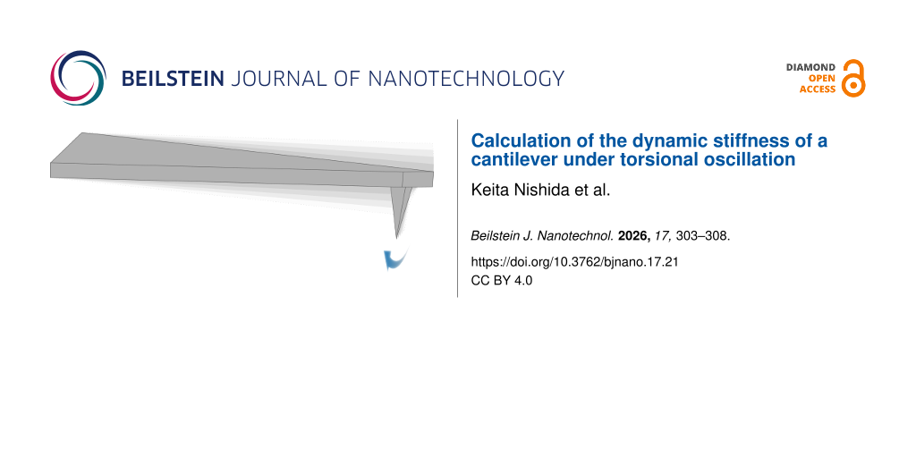
The photochemical pathway for ROS generation involves non-radiative relaxation of the excited PS, typically occurring through two primary mechanisms . Type-I reactions involve electron transfer, producing radical species such as superoxide anions, while type-II reactions involve energy transfer, generating singlet oxygen, the primary cytotoxic agent in PDT (Figure 1). Although nanostructured PSs are often associated with type-I reactions and molecular PSs with type-II reactions, the generation of one ROS type frequently triggers cascades leading to others, making the distinction primarily about the predominant mechanism rather than exclusivity.
![[2190-4286-17-27-1]](https://www.beilstein-journals.org/bjnano/content/figures/2190-4286-17-27-1.png?scale=2.0&max-width=1024&background=FFFFFF)
Figure 1: Photosensitizers in PDT: generations and types of PSs and their limitation, interaction of PSs with a cancer cell, mechanism of ROS generation by PSs, and induced death pathways (necrosis or apoptosis). The figure was created in BioRender. Syrocheva, A. (2026) https://biorender.com/mwpt7wl. This content is not subject to CC BY 4.0.
Efficacy and safety of PDT are largely determined by the PS’s properties . An ideal PS must exhibit a high absorption coefficient in the near-infrared (NIR) range to enable deep tissue penetration, addressing a key PDT limitation. However, the use of longer wavelengths (>800 nm) is not practical due to their inefficiency in exciting an oxygen molecule from the triplet ground state to the excited singlet state . A high quantum yield for ROS generation requires efficient excitation and minimal competing non-ROS-producing relaxation pathways. Low dark toxicity minimizes harm in the absence of light. High stability and low aggregation under physiological conditions are essential for maintaining photochemical activity . A balanced hydrophilicity/lipophilicity ensures selective accumulation in target tissues, such as tumors via the EPR effect, and efficient cellular internalization. Low photobleaching sustains activity during treatment, while biocompatibility and biodegradability reduce adverse effects and enhance the therapeutic index. PSs are classified into generations, each reflecting progressive improvement (Figure 1).
The first generation of PSs included hematoporphyrin derivatives (e.g., Photofrin) . The second generation of PSs encompasses purified compounds, including porphyrins , chlorins (e.g., chlorin e6 (Ce6) , mTHPC , bacteriochlorins , and phthalocyanines ), as well as non-porphyrinic structures like BODIPY dyes modified for enhanced ROS generation and natural compounds such as hypericin and curcumin, primarily used in antimicrobial PDT . Third-generation PSs focus on targeted delivery, achieved by conjugating second-generation PSs with carriers like antibodies, peptides, or nanoparticles to enhance specificity and reduce off-target effects .
The third generation of PSs should demonstrate minimal accumulation in healthy tissues and high selectivity toward pathological sites, thereby reducing side effects and increasing the therapeutic index. In this context, the use of nanocarriers for PS delivery gains particular importance, offering new opportunities to enhance both the selectivity and safety of PDT . Currently, nanoparticles serve as versatile delivery system for a broad range of molecular therapeutics, including small molecules , RNA , plasmids , CRISPR/Cas systems , and other bioactive agents.
Several key advantages of nanocarriers in PDT can be highlighted: (i) improved solubility and stability of PSs, (ii) enhanced delivery through both passive and active targeting mechanisms, (iii) controlled and stimuli-responsive release profiles, (iv) overcoming of tumor hypoxia, (v) efficient light conversion, and (vi) the development of multifunctional platforms by combining PDT with other therapeutic modalities . Additionally, nanoparticles enable theranostic applications by facilitating tumor visualization through various imaging techniques , including fluorescence imaging , computed tomography , and magnetic resonance imaging (MRI) .
The development of nanobiotechnology in PDT opens new horizons for creating more effective and safer cancer treatments. Further research in this field is expected to drive the design of personalized PDT approaches tailored to the unique characteristics of individual patients and tumors, as well as to broaden the clinical application of PDT across a wide range of oncological diseases.
Biomimetic nanoparticles: principles and platforms The BNP concept: nature-inspired deliveryAdvances in synthetic methods have enabled the fabrication of nanoparticles with a wide range of compositions, sizes, shapes, and surface properties . These tunable characteristics make nanoparticles highly versatile for applications in disease diagnosis and sensing technologies. Despite the promising potential of nanomedicine, significant progress has been limited. This can be attributed to the low targeted accumulation of the nanocarriers at the desired site of action. For instance, in cancer therapy, the accumulation of nanoparticles within solid tumors typically does not exceed 1% of the administered dose . The limited progress in achieving higher carrier targeted accumulation can be attributed to two main factors: (i) rapid clearance of the nanoparticles from the body, that is, nanoparticles are cleared from the systemic circulation by the organs and the cells of the mononuclear phagocyte system (MPS), primarily the liver and spleen, and (ii) ineffective overcoming of biological barriers, that is, nanoparticles have to overcome numerous biological barriers to reach the target tissue, including extravasation from the vasculature, penetration through the interstitial space, and cellular internalization at the target site . Conferring the carriers with a biomimetic shell that mimics the functions of biological cells potentially increases their circulation time and targeting properties, while significantly reducing the body’s immune response . BNPs utilizing a cell membrane to coat a synthetic core were reported in 2011. These nanoparticles consisted of a poly(lactic-co-glycolic) acid (PLGA) core coated with an outer layer derived from red blood cell (RBC) membranes . It was demonstrated that the presence of this biomimetic shell increased more than two times the circulation time of the carriers in the bloodstream compared to uncoated PEGylated (polyethylene glycol-coated) nanoparticles, representing the gold standard approach to nanoparticle circulation time. Since then, a wide variety of cell phenotypes have been explored as sources for biomimetic membranes to coat nanoparticles such that inherent biological functions and properties of specific cells could be exploited to enhance the performance and versatility of BNPs. According to some authors, biomimetic systems comprising proteins from cells to mimic their functions belong to the third generation of nanodelivery systems . The first generation of particles was based on surface modifications to reduce the interactions with immune cells and to increase biocompatibility. Among them, biocompatible polymers like PEG and PPE have been widely used . The second generation of nanocarriers harnessed surface functionalization with antibodies, peptides, and aptamers to increase the targeting of pathogenic tissues and cells by interacting with the receptors expressed on the surface of the target cells . In contrast, BNPs are designed to imitate the biological identity of cells through the corresponding surface proteins and clusters of biological molecules; these modifications enable the actions of aforementioned technologies and provide the particles with new functions.
Limitations of biomimetic nanoparticlesThe clinical translation of BNPs is primarily hindered by challenges related to large-scale production, reproducibility, and quality control. The multistep fabrication process, including extraction of cellular membranes and coating of nanoparticle cores, requires careful optimization to ensure stability and consistency .
Cell lysis is essential for the extraction of biomimetic membranes; the membrane composition is directly influenced by the severity of the lysis method. Common techniques include (i) ultrasound, that is, membrane rupture via pressure waves (20–50 kHz), requiring precise control to prevent heat damage ; (ii) freeze–thaw cycles, that is, cell disruption via narrow pores (enhanced at low temperatures) ; (iii) mechanical shearing, that is, cell disruption via narrow pores (enhanced at low temperatures) ; and (iv) osmotic pressure, that is, hypotonic solutions are used to induce swelling and rupture .
Lysis buffer selection (containing membrane disruptors and protease inhibitors) depends on cell type and target protein localization. For example, cancer cells and macrophages respond better to hypotonic protocols , and harsh homogenization suits cytomembrane protein extraction (above 30% cell composition).
Various methods exist for coating synthetic nanoparticle cores with biomimetic shells, each with unique advantages and limitations. A crucial factor is ensuring the correct “right-side-out” orientation of membrane proteins, which preserves system stability and enables proper interaction with target cells . Common techniques include (i) physical co-extrusion, where nanoparticles and membranes are forced through porous membranes to form uniform coatings; (ii) sonication, which uses ultrasonic energy to induce membrane fusion and often favors correct protein orientation; and (iii) electroporation, which creates temporary pores in membranes to facilitate coating while preserving protein integrity. Electroporation is simpler, more versatile, and scalable, especially when combined with microfluidic devices that allow for precise control over coating conditions, improving uniformity and reproducibility . Tailoring and combining these approaches enables the design of BNPs with desired functionalities, though challenges remain in standardizing protocols and scaling up production.
Other significant limitations include nonuniform coating, difficulties in purification, and maintaining sterility, especially for BNPs derived from bacterial membranes . Heterogeneous coverage can affect nanoparticle stability and drug delivery efficiency, while purification processes must effectively separate fully coated particles from impurities. For bacterial membrane-based BNPs, thorough detoxification is essential to eliminate toxic components without compromising immunogenic properties .
Finally, the biomimetic coating can influence drug release profiles, potentially hindering the controlled release of therapeutic agents . Optimizing these systems requires careful consideration of coating thickness, core material, and release mechanisms. Addressing these challenges through standardized production protocols, improved characterization, and enhanced biocompatibility will be critical to advancing BNPs toward clinical application.
Biomimetic nanocarriers, designed to replicate structure and functions of biological cells, present promising advantages for drug delivery and therapeutic applications. Nonetheless, they face notable challenges that require thorough investigation. Key areas of focus include developing standardized, scalable manufacturing techniques, elucidating their interactions with cells and mechanisms for crossing physiological barriers, enhancing stability for controlled drug release, and improving biocompatibility to ensure clinical safety. Tackling these challenges is essential to optimize biomimetic nanocarriers for better efficacy and successful translation into clinical practice.
Crossing of biological barriersBNPs face a series of biological barriers upon administration, which significantly influence their therapeutic and diagnostic effectiveness . One of the first challenges is opsonization, where plasma proteins form a “protein corona” on the nanoparticle surface, often reducing targeting specificity and biocompatibility. Coating nanoparticles with cell membranes, such as those from RBCs or leukocytes, can minimize protein corona formation, preserve targeting ligands, and extend circulation time by evading immune clearance. This strategy leverages natural “self-markers” like CD47 to inhibit phagocytosis and enhance passive tumor accumulation via the EPR effect .
Immune clearance by the MPS, primarily in liver and spleen, is another major hurdle. Biomimetic coatings help nanoparticles avoid rapid removal from the bloodstream, as demonstrated by prolonged circulation and increased tumor targeting in systems coated with erythrocyte or stem cell membranes . Such coatings can also facilitate homotypic targeting, where nanoparticles preferentially bind to and are internalized by cancer cells of the same origin, further improving specificity and reducing off-target effects.
The behavior of BNPs in the bloodstream is also shaped by hemodynamics and vessel wall interactions. Platelet-mimicking nanoparticles, for example, can adhere to vascular injury sites and inflamed tissues, enhancing accumulation at target areas and even supporting hemostasis. These properties are particularly valuable for treating inflammatory diseases and targeting tumors.
Crossing the blood–brain barrier (BBB) remains a significant challenge for nanomedicine. BNPs coated with membranes from cells capable of BBB penetration, such as neutrophils or cancer cells, show improved brain delivery . Additional modifications with targeting peptides can further enhance BBB crossing, opening new possibilities for treating neurological diseases.
Finally, successful therapy depends on efficient cellular uptake and drug release. Biomimetic shells can increase nanoparticle internalization through receptor-mediated endocytosis and homotypic binding, as well as improve colloidal stability. Strategies such as incorporating pH-sensitive components or lysosomal escape mechanisms further enable controlled drug release within target cells. Despite these advances, optimizing BNP design to overcome all biological barriers remains an active area of research essential for clinical translation.
BNP functionalizationThe development of biomimetic drug delivery systems has advanced rapidly, with multifunctional platforms emerging as a key focus . By leveraging the natural properties of cell membranes, such as immune evasion, prolonged circulation, and specific targeting, BNPs can be further enhanced through surface modifications to improve their therapeutic performance. These modifications enable BNPs to address complex challenges in drug delivery, including targeted transport, controlled release, and overcoming biological barriers.
One promising strategy is the creation of hybrid biomimetic coatings, where membranes from multiple cell types are fused to combine their unique functionalities. For example, NPs coated with a mix of RBC, cancer cell, and immune cell membranes can simultaneously evade the immune system, target tumors, and deliver drugs to metastatic sites. This approach has demonstrated improved blood circulation, enhanced tumor targeting, and increased therapeutic efficacy in preclinical models. However, the process of membrane fusion and the need for additional purification steps add complexity to manufacturing .
Genetic modification of source cells offers another avenue for BNP functionalization. By engineering cells to express specific targeting proteins or receptors before membrane extraction, it is possible to produce BNPs with highly selective targeting capabilities. For instance, membranes from CAR-T cells or stem cells engineered to overexpress homing receptors can be used to create BNPs that efficiently recognize and bind to tumor cells or inflamed tissues, minimizing off-target effects and systemic toxicity .
Post-synthesis surface modification is also widely used to enhance BNP functionality . Non-covalent methods, such as lipid insertion, allow for the integration of targeting ligands, peptides, or imaging agents into the membrane without disrupting protein activity. Covalent modifications, including the attachment of antibodies or peptides via amide or thiol–maleimide linkages, provide stable and specific functionalization but may risk altering membrane protein function if not carefully controlled. Enzymatic approaches can offer greater specificity but require additional steps and conditions.
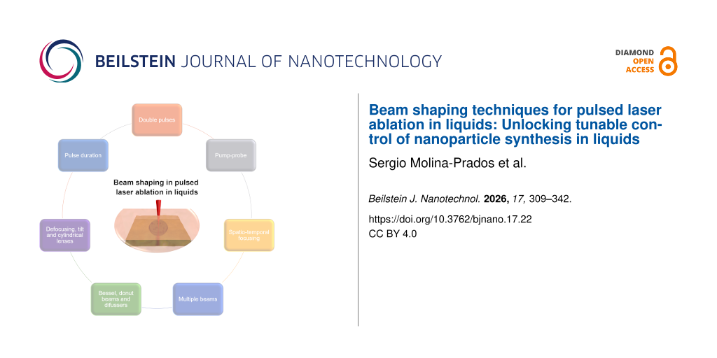
These advanced BNPs can be engineered to respond to specific stimuli, such as pH, enzymes, or external triggers like light, enabling controlled drug release at the target site . For example, pH-sensitive lipids or peptides can be incorporated to accelerate drug release in the acidic tumor microenvironment. Additionally, BNPs can be loaded with imaging agents or therapeutic enzymes to facilitate tumor visualization and penetration, further expanding their utility in cancer therapy. Targeting strategies can be further refined by integrating ligands such as arginylglycylaspartic acid (RGD) peptides, antibodies, or aptamers onto the BNP surface, enhancing their ability to recognize and bind to specific cell markers . Incorporating imaging dyes or contrast agents into the BNP membrane also enables real-time tracking and monitoring of drug delivery in vivo. The main stages of biomimetic nanoparticle fabrication and subsequent surface modification are summarized in Figure 2.
![[2190-4286-17-27-2]](https://www.beilstein-journals.org/bjnano/content/figures/2190-4286-17-27-2.png?scale=2.0&max-width=1024&background=FFFFFF)
Figure 2: Processes for fabricating and functionalizing biomimetic nanoparticles (BNPs), highlighting key methodologies for cell membrane extraction, coating, and surface engineering to achieve enhanced tumor targeting and therapeutic performance in photodynamic therapy. The figure was created in BioRender. Syrocheva, A. (2026) https://biorender.com/ksvnysj. This content is not subject to CC BY 4.0.
BNPs for enhanced delivery and specificity in PDTBNPs achieve spatial specificity within the TME through a hierarchical targeting strategy, combining passive accumulation mechanisms with innate or engineered active targeting capabilities. This multilevel approach significantly enhances the delivery efficiency of PSs compared to conventional nanocarriers .
Passive targeting primarily exploits EPR effect, a pathophysiological feature of many solid tumors characterized by leaky vasculature and impaired lymphatic drainage . BNPs amplify passive accumulation through their extended circulation time, a direct consequence of immune evasion conferred by their biomimetic coatings . Prolonged systemic exposure increases opportunities for extravasation through tumor vasculature. For instance, erythrocyte membrane-coated BNPs consistently demonstrate 1.5- to 2.0-fold higher tumor accumulation than their uncoated counterparts with identical cores, attributable solely to superior circulation half-life and EPR potentiation . While EPR is a universal mechanism for nanocarriers, the significant enhancement provided by biomimetic coatings, particularly erythrocyte and mesenchymal stem cell membranes, represents a critical advantage over synthetic platforms, enabling greater passive PS deposition within tumors .
Active targeting utilizes specific molecular interactions to enhance tumor cell binding and internalization. BNPs employ both innate (source cell-derived) and engineered strategies to achieve this. Innate active targeting exploits the inherent biological tropism preserved on the source cell membrane, requiring no additional modification . As exemplified by mesenchymal stem cell (MSC) membrane-coated BNPs, this system can utilize the natural homing capability of the source cells (e.g., MSCs migrating towards TME chemokine gradients like SDF-1α and CCL2) . This conserved tropism results in significantly higher tumor accumulation compared to untargeted controls .
The biological specificity of different cell types is determined by proteins present on their plasma membranes, which not only serve structural roles but also mediate cell–cell adhesion, either homotypic or heterotypic, depending on molecular affinity. The primary mechanisms underlying homotypic binding involve various classes of adhesion proteins, such as integrins, selectins, cadherins, and members of the immunoglobulin superfamily (IgSF). Integrins typically interact with the extracellular matrix, whereas selectins, cadherins, and IgSF proteins are primarily responsible for cell–cell adhesion . The cytoplasmic and extracellular domains of these proteins confer distinct physicochemical properties that influence cellular interactions and tissue organization. Given the crucial role of integrins in tumor progression, there is significant interest in their use not only as markers of malignant potential but also as targets for precision therapies.
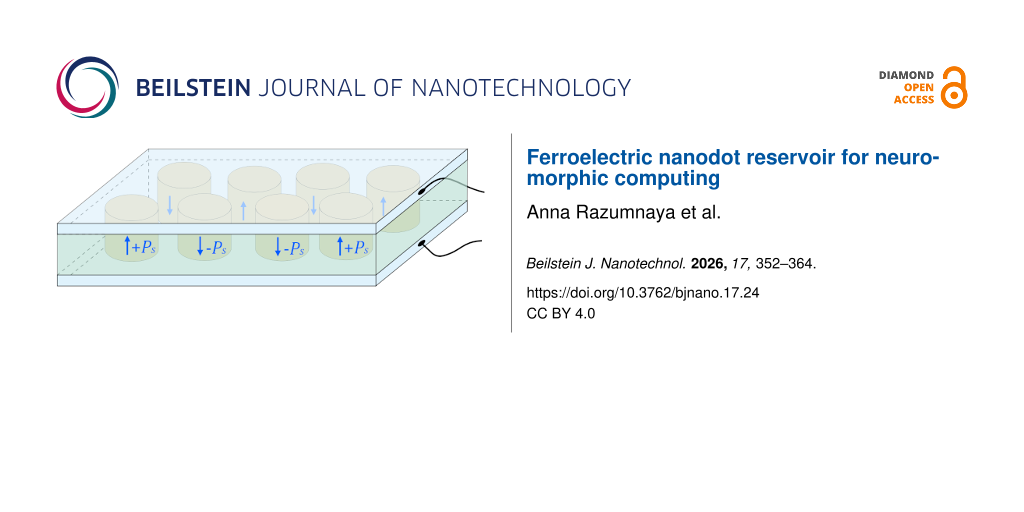
One of the most extensively studied integrins, αVβ3, is a transmembrane receptor for extracellular matrix proteins that binds cyclic RGD peptides with high affinity and interacts with SRC oncogene to promote lymph node metastasis. For example, RGD-coated BNPs enhanced doxorubicin (DOX) uptake by 1.4 times in αVβ3-positive U-87 MG glioblastoma cells compared to αVβ3-negative HeLa cells . In urological cancers, integrin α5β1 is upregulated, facilitating cell adhesion to fibronectin; PHSCN peptide-targeted BNPs have shown efficacy in inhibiting prostate cancer metastasis in preclinical and clinical settings . High malignancy cancer cells display reduced homotypic adhesion but increased heterotypic adhesion to endothelial cells, mediated by molecules such as IL6R , CXCR4 , and EPCAM , which are targeted via peptide-functionalized BNPs to inhibit metastasis. Additionally, NGR peptides targeting aminopeptidase N have improved photoinduced activity of chlorin e6 in fibrosarcoma cells ; dual peptide modification of PLGA nanoparticles enhanced brain targeting in glioma models . Integrin expression varies across tumor types, influencing homotypic targeting strategies . A membrane coverage of 50% and more enables cellular uptake of individual nanoparticles, while lower coverage requires aggregation for internalization . Using cancer cell membranes (CCMs) with high coverage on BNPs ensures selective targeting through integrin-mediated homotypic binding, promoting efficient therapeutic delivery (Figure 3). Relevant proteins enabling homotypic targeting via BNPs are listed in Table 1.
![[2190-4286-17-27-3]](https://www.beilstein-journals.org/bjnano/content/figures/2190-4286-17-27-3.png?scale=2.0&max-width=1024&background=FFFFFF)
Figure 3: Selective targeting and internalization via homotypic binding and receptor-mediated endocytosis of biomimetic nanoparticles coated with cancer cell membranes, enabling enhanced photosensitizer accumulation and reactive oxygen species generation for improved photodynamic therapy. The figure was created in BioRender. Syrocheva, A. (2026) https://biorender.com/jjvlem9. This content is not subject to CC BY 4.0.
Table 1: A list of proteins that can be incorporated into biomimetic nanoparticles to facilitate targeted delivery via homotypic binding.
Protein Cell lines Cancer type Application in drug delivery Ref α- and β-integrins 4T1, BT-20, B16, C4-2B breast cancer, breast carcinoma, melanoma, prostate cancer, bone metastases In biomimetic nanoparticles, proteins and peptides containing the RGD (arginine-glycine-aspartate) motif are most often used for targeted binding to α- and β-integrins. The main proteins used in such systems are fibronectin, vitronectin, and laminin, as well as synthetic RGD-based peptides (for example, cyclic RGD peptides). The use of fibronectin in the coating of nanoparticles loaded with doxirubicin, tannic acid, and iron contributed to the targeted therapy of the B16 tumor with high expression of integrin a5b3. Intracellular absorption of nanoparticles was confirmed by confocal laser scanning microscopy. For the treatment of prostate cancer C4-2B, poly(lactic-co-glycolic acid) nanoparticles coated with cancer cell membranes containing integrin β3, a subunit of integrin α5ß3, were used. The resulting nanoparticles demonstrated a fourfold increase in absorption in C4-2B cells compared to uncoated nanoparticles, which was measured using flow cytometry. E-cadherin and N-cadherin 4T1 cells breast cancer, lung metastases 4T1 cancer cell membrane-coated paclitaxel nanoparticles showed 3.3 times and 2.5 times higher accumulation in primary breast tumors and lung metastases versus uncoated NPs. Negative controls (RBC-coated and liposome-coated NPs) had lower uptake. E- and N-cadherins mediated binding. P-selectin H22 cells hepatoma Platelet membrane-coated biomimetic nanoparticles loaded with bufalin targeted hepatoma via P-selectin–CD44 interaction, enhancing uptake and inhibiting tumor growth. E-selectin A549, Hs 578T circulating tumor cells (various) Surfactant–nanotube biomimetic complexes with E-selectin facilitated capture of circulating A549 and Hs 578T carcinoma cells, confirmed by flow cytometry. galectin-1 and galectin-3 4T1 cells breast cancer biomimetic nanoparticles containing calcium phosphate and STING Mn2+ agonist, Cypate photosensitizer, and indolamine 2,3-dioxygenase 1 (IDO1); the inhibitor was coated with galactose to act on galectin-1 and galectin-3 receptors; after intravenous administration to mice, this system demonstrated high accumulation in 4T1 tumors, which ensured accurate diagnosis and effective elimination.Early studies on BNPs often employed simple coatings derived from CCMs. This approach effectively camouflaged the nanoparticles by conferring the properties of “self” cells, thereby enhancing biocompatibility and reducing neutrophil-mediated immune clearance. For instance, BNPs loaded with DOX and coated with glioblastoma cell membranes were employed as a targeted therapy for malignant tumors. The presence of surface adhesion molecules, including N-cadherin, galectin-3, and epithelial cell adhesion molecules, facilitated enhanced uptake of the drug in the human glioblastoma U87 MG cell line. This resulted in an 8.5-fold increase in cellular internalization compared to uncoated nanoparticles . However, over time, it became evident that such simple membrane coatings have limited functional capabilities. This realization has driven the development of additional surface modifications aimed at improving targeting specificity to particular cells or tissues, enhancing stability in the bloodstream, controlling drug release kinetics, and tailoring nanoparticle systems to meet diverse clinical requirements. Recent advances in this field have demonstrated the remarkable efficacy of homophilic targeting. Specifically, the involvement of CD47 from lung CCM has been shown to facilitate evasion of phagocytosis, while N-cadherin, galectin-3, CD44, and CD326 mediate homotypic binding to cancer cells; this resulted in a threefold increase in the accumulation of PLGA nanoparticles within the tumor . A notable study aimed to evaluate the translational potential of biomimetic delivery systems for precise tumor therapy. Gold@carbon nanoparticles were coated with proteins derived from different cell types, including a squamous cell carcinoma cell line and patient-derived cells. To address tumor heterogeneity, these systems were tested across various xenograft models, namely, cell line-derived subcutaneous xenografts, orthotopic tongue xenografts, immunocompetent primary and distant tumor models, and patient-derived xenografts. In all models, the biomimetic coating enhanced nanoparticle accumulation within tumors and improved therapeutic efficacy. The authors emphasize the importance of such comprehensive evaluations to facilitate the translation of these findings into clinical practice .
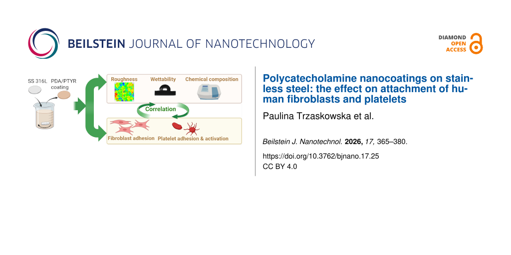
A prominent trend in drug delivery is the creation of multifunctional, stable, and biocompatible platforms with tunable biological activity, often achieved by using hybrid membrane coatings that combine proteins from multiple cell phenotypes, thereby enhancing their therapeutic and diagnostic versatility. The concept of hybrid membrane coating was first introduced in 2017 , where PLGA nanoparticles were coated with combined erythrocyte and platelet membranes, leveraging their unique functions to evade immune clearance and target inflammatory sites. Since then, an increasing variety of differentiated cell membranes have been combined to modify biomimetic nanoparticle surfaces, such as cancer cell–erythrocyte and macrophage-–cancer cell hybrids (Figure 4). For combinational therapy of triple-negative breast cancer, BNPs based on tumor cell membranes and PLGA loaded with the cytolytic peptide melittin and the photosensitizer mTHPC have been developed. In vitro and in vivo studies demonstrated that these nanoparticles mitigated the acute toxicity of melittin and enhanced tumor accumulation of mTHPC in 4T1 tumor-bearing mice, while facilitating accelerated melittin release due to disruption of the membrane integrity .
![[2190-4286-17-27-4]](https://www.beilstein-journals.org/bjnano/content/figures/2190-4286-17-27-4.png?scale=2.4&max-width=1024&background=FFFFFF)
Figure 4: Modifications of biomimetic nanoparticle coatings using erythrocyte, macrophage, and cancer cell membranes, conferring unique functional properties including prolonged systemic circulation, immune response evasion, and homotypic tumor targeting for enhanced photodynamic therapy. The figure was created in BioRender. Syrocheva, A. (2026) https://biorender.com/ffuwoj1. This content is not subject to CC BY 4.0.
Similar to other nanodelivery systems, a wide variety of surface functionalization strategies have emerged to enhance targeting efficiency in BNPs. In a 2022 study, a sophisticated biomimetic system was developed comprising paclitaxel nanocrystals coated with SKBR-3 cell membranes. The membrane coating was further functionalized with Herceptin, a monoclonal antibody that selectively binds to HER2 receptors, rendering the platform a promising candidate for the treatment of HER2-positive breast cancer . In vivo experiments demonstrated homotypic targeting, significantly enhanced cellular uptake compared to uncoated nanocrystals, and confirmed the system’s ability to induce apoptosis in HER2-positive breast cancer cells. Another study reported the development of a multifunctional biomimetic system coated with erythrocyte membranes for the co-delivery of chlorin e6 and DOX to enhance synergistic targeted tumor therapy. This system exhibited high targeting efficiency toward HepG2 cells via folic acid receptor-mediated endocytosis, followed by effective tumor eradication through singlet oxygen generation. Modification with erythrocyte membranes also conferred prolonged circulation time to the nanoparticles, attributed to the presence of the CD47 molecule .
An alternative approach to the functionalization of biomimetic coatings is based on genetic engineering. In a recent study employing this strategy, the cell membrane from a genetically engineered HEK-293 cell line expressing synthetic SpyCatcher proteins was used to coat NPs, enabling targeted binding to both tumor cells and viral particles. Treatment with these nanoparticles in a mouse model of ovarian cancer suppressed tumor growth and improved survival outcomes . For the therapy of uveal melanoma, BNPs coated with recombinant low-density lipoproteins were developed to deliver the PS verteporfin and the vascular-normalizing agent dexamethasone. Following intravenous administration, these nanoparticles selectively accumulated in tumor tissue; verteporfin generates ROS, resulting in tumor cell death, while the ROS also facilitated rapid release of dexamethasone, which inhibited neovascularization in the tumor microenvironment. Thus, the ability to program specific protein sequences allows for their precise display on the nanoparticle surface in physiologically relevant conformations and orientations, thereby enhancing their functional activity in binding to cellular receptors.
A key emerging trend in such delivery systems is the development of smart platforms that respond to external stimuli, enabling remote-controlled release of therapeutic payloads. A significant advancement came from Wang’s group in 2023 , who developed “adaptive” CCM-coated nanoparticles that could dynamically alter their surface properties in response to the TME. By incorporating pH-sensitive linkers between the membrane coating and targeting ligands, these nanoparticles maintained their stealth properties during circulation but exposed their homophilic binding sites specifically within the acidic tumor environment, resulting in a 2.5-fold increase in tumor-specific uptake compared to conventional membrane-coated systems.
Main classes of biomimetic nanoplatforms for photosensitizer deliveryExosome-based platforms utilize naturally secreted extracellular vesicles (40–100 nm) possessing a lipid bilayer and a unique surface protein profile derived from various cell types (e.g., dendritic cells, T cells, and macrophages) . Their endogenous role in intercellular communication confers low immunogenicity and high biocompatibility, facilitating evasion of immune clearance . A key advantage is their ability to penetrate biological barriers, including the BBB , enabling access to challenging tumor sites like glioblastoma . The surface-exposed proteins support inherent cell-specific interactions, which can be augmented by engineering targeting ligands for improved tumor specificity . In PDT, exosomes effectively deliver PSs like Ce6, demonstrating enhanced tumor accumulation and photodynamic efficacy compared to free PSs . Challenges include complex isolation procedures, heterogeneity, scalability limitations, and potential loss of membrane protein activity during processing .
CCM-coated platforms involve cloaking synthetic nanoparticle cores (e.g., PLGA, silica, and metal-organic frameworks (MOFs)) with membranes derived from tumor cells . This biomimetic strategy primarily exploits homotypic targeting; the CCM retains adhesion molecules (e.g., galectin-3 or T-antigen binding proteins) enabling preferential binding and deep penetration into homologous tumor tissues. CCM-coated NPs have successfully delivered diverse PSs (e.g., Ce6 , PCN-224 , and IR-780 ), often in combination therapies (e.g., chemo-PDT and immune checkpoint blockade ), achieving high tumor accumulation and potent PDT responses in preclinical models. Critical limitations include the inherent specificity of homotypic targeting to the parent cancer cell type, restricting broad applicability, and scalability challenges in membrane production and fusion.
RBC membrane-coated platforms employ the readily available membranes of RBCs to encapsulate nanoparticle cores . Their primary advantages are their exceptional ability to evade the immune system and their long circulation half-life, which exceeds 100 days in vivo for native RBCs . These properties have popularized two major strategies, that is, using RBC membranes as a biomimetic shell for nanoparticles and directly targeting circulating RBCs in vivo for drug delivery . This is largely mediated by CD47 expression on the RBC membrane, which delivers a potent “do not eat me” signal by binding to SIRP-α on phagocytes . This inherent property ensures high biocompatibility and minimal toxicity. While lacking innate tumor targeting, RBC-coated NPs can be effectively functionalized with ligands (e.g., folate or peptides like RGD ) to achieve active targeting, enhancing accumulation at tumor or infection sites. These platforms efficiently deliver PSs (e.g., Ce6 , TPC , MC540 , indocyanine green (ICG) , or IR-780 ), leveraging the EPR effect, long circulation times, and potential ligand-mediated targeting to improve tumor PS levels, thereby enhancing PDT efficacy and enabling combination strategies (e.g., with anti-PD-1 immunotherapy ). The need for additional functionalization for specific targeting and potential batch variability in membrane extraction represent key considerations.
MSC membrane-coated platforms utilize membranes derived from stem cells to cloak nanoparticle cores . A defining characteristic is their ability to mimic the tumor tropism of native MSCs. This is achieved through the retention of chemokine receptors (e.g., CXCR4) on the MSC membrane, which mediate specific adhesion to ligands (e.g., SDF-1α) prevalent on the endothelium within TME . This property facilitates active migration towards tumor and metastatic sites. Additionally, MSC membranes confer low immunogenicity and reduced MPS clearance, enhancing overall biocompatibility and tumor penetration. MSC-coated NPs have been employed to deliver PSs such as Ce6 , ZnPc, and MC540 , demonstrating improved tumor accumulation and cellular uptake compared to non-biomimetic counterparts. While the tropism is advantageous, the precise molecular mechanisms governing MSC homing require further elucidation, and scalability/reproducibility of MSC culture and membrane isolation remain practical challenges.
Immune cell membrane-based platforms incorporate membranes from various immune cells (e.g., macrophages, neutrophils, dendritic cells (DCs), T cells, and natural killer (NK) cells) onto nanoparticle cores or utilize derived extracellular vesicles . Immune cell membrane-based BNPs represent a breakthrough platform that seamlessly integrates PDT with immunotherapy. The membranes retain source cell-specific functionalities: (i) Macrophage membranes exhibit tropism towards inflammatory sites and tumors via chemokine receptors (e.g., CCR2) and adhesion molecules. Pre-polarization to an M1 phenotype ex vivo can further equip these nanoparticles to reprogram the immunosuppressive TME . (ii) Neutrophil membranes inherently target inflamed endothelium and tumor sites; facilitate crossing vascular barriers . (iii) DC membranes possess inherent homing to lymph nodes and carry tumor-associated antigens, promoting antigen presentation and T-cell activation when combined with PDT-induced ICD . (iv) T-cell membranes (including CAR-T) enable specific antigen recognition via TCR or CAR . (v) NK cell membranes inherit natural cytotoxicity receptors (e.g., NKG2D and NKp30) for innate tumor recognition and killing activation . (vi) Platelet-derived membranes exhibit natural adhesion to damaged vasculature (common in tumors) and circulating tumor cells (CTCs); they also express CD47 for extended circulation and modulate immune cell interactions and inflammation .
These properties allow for deep tumor penetration and direct interaction with immune components within the TME. Consequently, immune cell BNPs can deliver PSs while simultaneously modulating the immune landscape; for instance, they promote ICD via PDT and concurrently deliver checkpoint inhibitors (e.g., anti-PD-1/PD-L1) or immunomodulators (e.g., R848). This potent combination therapy approach is explored further in the context of immune modulation. Challenges include the complexity and variability of immune cell membrane isolation, potential for immune rejection depending on allogenicity, reproducibility, and scalability. Hybrid platforms represent an advanced strategy where membranes from two or more distinct cell types (e.g., RBC + cancer cell, RBC + immune cell, and cancer cell + macrophage) are fused to create a single biomimetic coating . This approach combines the unique advantages of each parental membrane.
PS loading into biomimetic nanocarriersEfficient encapsulation of PSs into BNPs is a pivotal step that directly influences the overall therapeutic outcome of PDT. The success of PDT hinges not only on the intrinsic photochemical properties of the PS but also on its effective delivery to the tumor site, stability during circulation, bioavailability within the TME, and ability to be activated by light to generate ROS. BNPs, by virtue of their biological surface functionalities and synthetic nanoscale architecture, offer sophisticated platforms that maximize these criteria. However, the process of loading PS into such carriers must be carefully optimized to maintain the delicate balance between high loading capacity, preserved photophysical activity, controlled release, and targeting specificity. Several advanced strategies have been developed and tailored to integrate PSs into BNPs. These approaches take into consideration the hydrophobic nature of the PS, the chemical functionalities available for conjugation, and the physicochemical characteristics of the carrier. For instance, encapsulation techniques must ensure that PS molecules remain stably entrapped within the nanoparticle core to prevent premature leakage during blood circulation while enabling efficient release or activation within the tumor site.
A widely adopted approach is to first encapsulate hydrophobic PS molecules into synthetic nanoparticle cores. These stable PS-loaded cores are subsequently cloaked with natural cell membranes, imparting immune evasion, prolonged circulation, and targeting. Ce6 was loaded to methoxy PEG-PLGA NPs via microfluidic flow, covered with RBC membrane and modified with folic acid. The loading efficiency was 20%, and coating with the biomimetic membrane reduced the initial burst release during the first 4 h while sustaining gradual release over 48 h. The biomimetic coating increased the rate of cancer cell apoptosis induced by the photodynamic effect by 50% and prolonged the circulation time of the nanoparticles in the bloodstream .
BNPs composed of materials such as melanin or polydopamine exploit strong π–π stacking interactions with aromatic PSs like Ce6 to achieve high loading capacity. For instance, melanin-based nanoparticles loaded with Ce6 (loading efficiency ≈ 30%) showed enhanced biocompatibility and water solubility, with synergistic photodynamic and photothermal effects under imaging guidance .
Another strategy involves chemically attaching porphyrin-based PSs bearing conjugatable functional groups (e.g., carboxyl) onto the surface of bioinspired nanoparticles such as noble metal nanoparticles (Ag and Au). Stable ester bond formation secures the PS to the nanocarrier surface, offering hydrophilicity, biocompatibility, and enhanced biological delivery. Loading efficiencies of ≈16% have been reported for synthesized porphyrin PS and bioinspired metal nanoparticles (rich in –OH groups), suitable for dual PDT and photothermal therapy (PTT) .
Some biomimetic platforms enable co-loading of the PS with other agents to boost therapeutic outcomes. For example, DOX was preloaded into RBC nanovesicles while Ce6 was encapsulated in polymeric cores coated with RBC membranes . Doxorubicin and ICG were loaded to BNPs with CCMs and in vivo demonstrated high tumor ablation with low systemic toxicity. This multifunctional system combines chemotherapy and PDT, together with a number of diagnosis tools .
Beyond efficient loading, controlled PS release from BNPs represents a complementary critical factor governing PDT efficacy within the TME. Biomimetic coatings not only facilitate high payload delivery but also enable stimuli-responsive release mechanisms tailored to TME conditions, including light-triggered bursts (e.g., >70% PS liberation within 5 min via azobenzene linker cleavage under 660 nm irradiation ), enzyme-sensitive untethering (MMP-2 hydrolysis of peptide linkers overexpressed in tumor ECM ), and biomimetic pH gradients (sustained zero-order release over 24–72 h during endosomal acidification ). These strategies ensure spatiotemporal ROS generation while minimizing off-target phototoxicity, as exemplified by membrane-cloaked systems exhibiting reduced initial burst (<20% at 4 h) and prolonged therapeutic windows. These integrated loading/release optimizations are illustrated in Figure 5.
![[2190-4286-17-27-5]](https://www.beilstein-journals.org/bjnano/content/figures/2190-4286-17-27-5.png?scale=2.0&max-width=1024&background=FFFFFF)
Figure 5: Biomimetic nanoparticle strategies for PS loading and controlled release in PDT. (A) Loading methods; (B) controlled-release triggers; (C) quantitative benchmarks for loading and release estimation. The figure was created in BioRender. Syrocheva, A. (2026) https://biorender.com/cknfimy. This content is not subject to CC BY 4.0.
In summary, biomimetic platforms coated with cell membranes or molecules offer a highly promising strategy for the targeted delivery in cancer theranostics, particularly in PDT. By leveraging homotypic recognition and binding to tumor cells, these coatings enhance selective accumulation within tumors while minimizing adverse effects on healthy tissues. Moreover, the integration of diverse therapeutic agents within these BNPs can yield synergistic antitumor effects, thereby improving the overall efficacy of PDT. This approach represents a significant advancement in the development of precise and effective cancer treatments. The precise loading and controlled release of PS into BNPs represent equally pivotal determinants of PDT efficacy, warranting comprehensive analysis alongside encapsulation strategies. While high loading capacities ensure adequate PS payloads for sufficient ROS generation at tumor sites, release kinetics govern bioavailability within the tumor microenvironment, spatiotemporal control of photoactivation, and minimization of off-target phototoxicity during systemic circulation.
Addressing tumor hypoxia: BNP innovationsThe intrinsic hypoxic nature of solid tumors poses a major challenge to PDT efficacy, as limited oxygen availability restricts ROS production. Furthermore, the oxygen consump
Comments (0)