Characterization of Hyrtl’s anastomosis: anatomical types, measurements and fetoplacental correlation
Purpose
Hyrtl’s anastomosis, a vascular connection between the umbilical arteries close to its placental attachment that plays a crucial role in maintaining balanced fetoplacental circulation. Despite its clinical implications, its morphological variations remain underexplored.
Methods
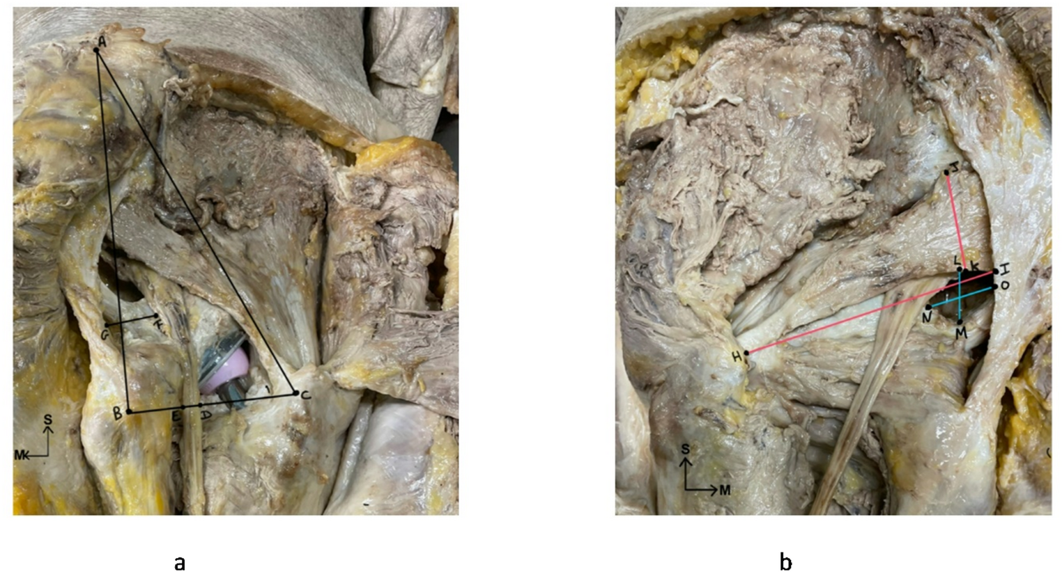
Term placentas collected after delivery were analysed for the presence, type, and location of Hyrtl’s anastomosis. Gross dissection and morphometric analysis were done. The distance between Hyrtl’s anastomosis and cord insertion was measured.
Results
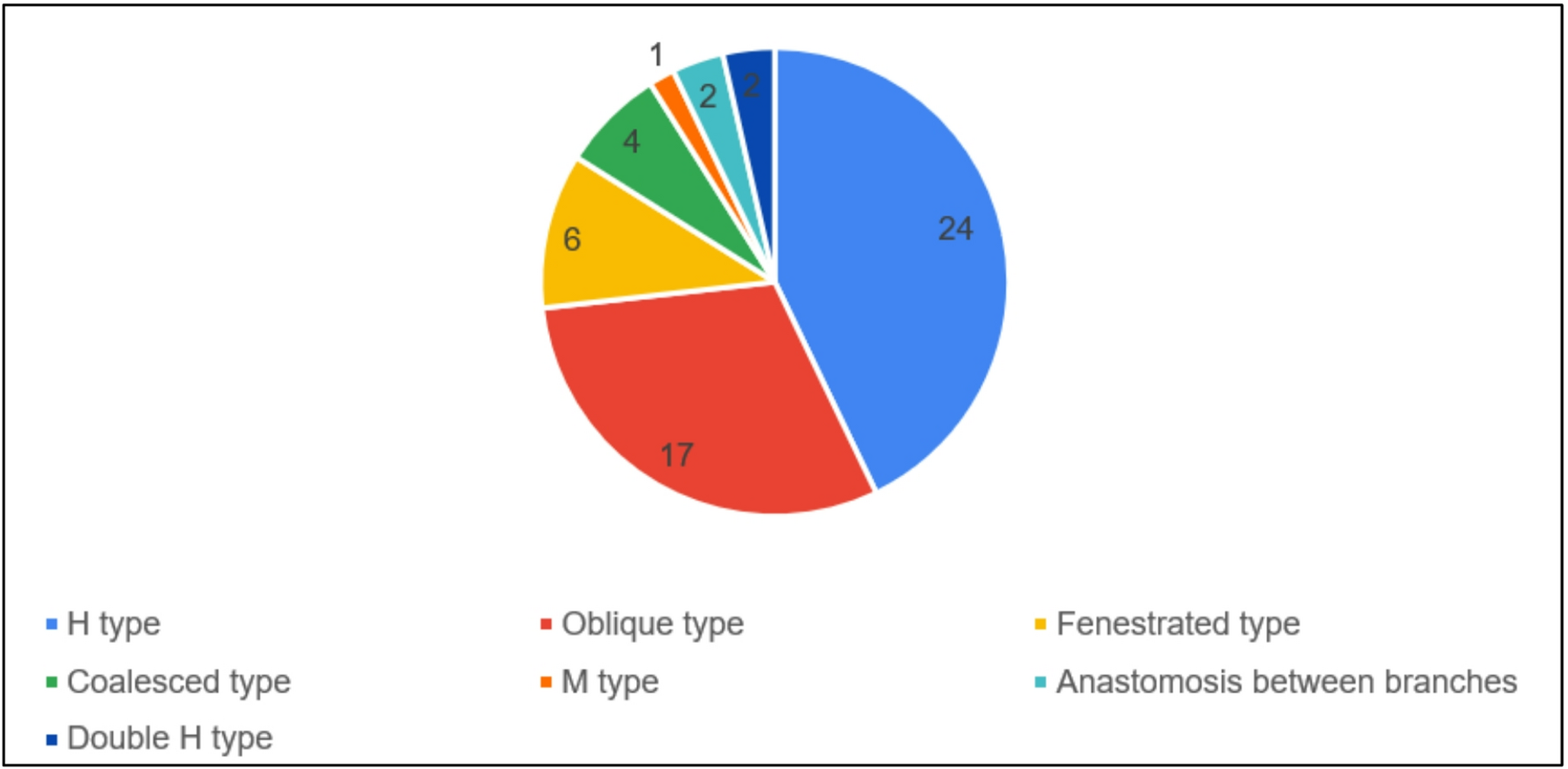
Hyrtl’s anastomosis was observed in 56 of 111 placentas (50.5%). Most cases demonstrated a single anastomosis (91.1%), while multiple anastomoses were observed in 8.9%, including two cases of a rare double H-type. The H-type was the most frequent morphological pattern (42.9%), followed by oblique (30.4%), fenestrated (10.7%), coalesced (7.1%), M type (1.8%), and anastomosis between branches (3.6%). The mean distance of Hyrtl’s anastomosis from cord insertion was 14.3 ± 10.2 mm (range: 1.3–70 mm). No significant differences were found in maternal, placental, or neonatal outcomes between groups; however, placental weight showed a stronger correlation with neonatal weight when Hyrtl’s anastomosis was present, suggesting subtle efficiency advantages.
Conclusion
Morphological heterogeneity in Hyrtl’s anastomosis may affect fetoplacental hemodynamics. Recognising such anatomical variances emphasises the significance of including Hyrtl’s anastomosis assessment into routine antenatal ultrasonography, demonstrating the clinical utility of anatomical knowledge.


Comments (0)