Tracheal morphometry using computed tomography in North Indian adults without any respiratory illness and its correlation with spirometry indices
Purpose
The data regarding the tracheal morphometry in India, especially in vivo setting in healthy adults, is scarce. The current study aimed to assess tracheal morphometry in the North Indian population using computed tomography (CT) scans and to explore its potential impact on pulmonary functions.
Methods
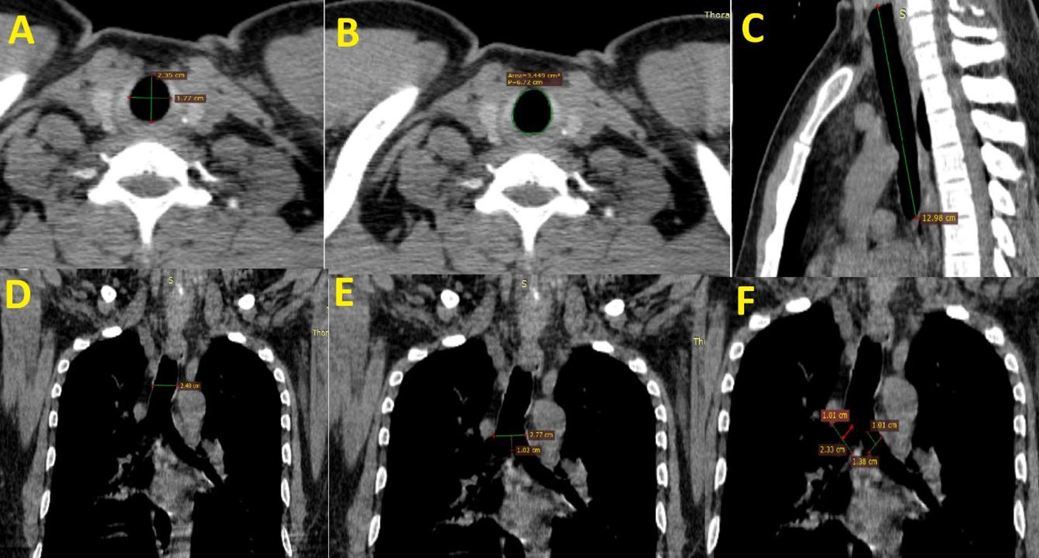
The study was a cross-sectional, observational design conducted in a north-Indian tertiary-care hospital. Participants included adults (> 18 years) undergoing CT thorax for non-respiratory symptoms. Exclusion criteria included significant smoking history, abnormal lung parenchyma, or prior thoracic surgery. Tracheal measurements (length, transverse diameters, and anteroposterior diameter) were taken. Pulmonary function was assessed with spirometry, focusing on Forced-Vital-Capacity (FVC), Forced-Expiratory-Volume (FEV₁), and the FEV₁/FVC ratio.
Results
The mean age of the study population was 60.21years, and the majority were males (82.9%). The study found significant variation in tracheal dimensions across participants, with the tracheal diameter at various levels: 1.70 cm at the C7 vertebra, 1.84 cm at the upper border of the arch of the aorta, and 2.16 cm at one centimeter above the carina. The trachea had a consistent length of 13.43 cm across the population. Tracheal dimensions showed significant differences based on sex, with males having larger diameters. However, only the diameter of the left main bronchus showed a weak correlation with the FEV₁/FVC ratio.
Conclusion
This study provides essential baseline data on tracheal morphometry in the North Indian population. The lack of correlation between tracheal dimensions and pulmonary functions suggests that while anatomical variations are prevalent, they may not directly impact lung function in a clinically significant manner.


Comments (0)