
Remember me




An injury to the central nervous system can lead to the loss of upper motor neuron inhibitory pathways and altered intra-spinal afferent signaling. This alteration causes spinal reflex hyperexcitability hallmarked by a velocity-dependent abnormal stretch response with increased motor tone and muscle activation known as spasticity. It is important to note that this upper extremity spasticity is one component of upper motor neuron syndrome, the others including motor weakness, easy fatigability, lack of cortical control or neglect, co-contraction, and dysynergistic motor patterning. We focus on spasticity as it is a feature that is more readily alleviated by surgical intervention but understanding these other features when evaluating patients and formulating treatment plans is paramount to setting expectations and understanding the complexity of the problem at hand.
The most common etiologies for upper motor neuron syndrome are cerebral palsy (CP), traumatic brain injury (TBI), cerebrovascular incidents (CVA), and spinal cord injury (SCI). The rates of long-term spasticity in the CVA population alone are reported at 30–60%, with potential higher numbers in TBI patients [1]. With growing evidence that surgical intervention can alleviate symptoms in these populations, it is currently estimated that less than 1% of these patients are ever managed with surgery [2]. It is therefore of the goal of this article to summarize the current evidence of microsurgical applications in the treatment of spasticity, improve awareness of what surgery can offer to both surgeons and rehabilitation teams, and ultimately advocate the utilization of hyperselective neurectomies (HSN), tendon lengthenings and transfers to improve the lives of these patients.
Non-Surgical TreatmentPhysical and occupational hand therapy, botulinum toxin, splinting, casting may all be employed as non-surgical modalities in the acute setting. Generally, this timeline represents about 12 months after CVA/SCI, and 15–18 months in the TBI population to gain the majority of motor recovery that is possible [3]. In addition, most data suggests spasticity after this time period does not sporadically resolve. It is important to immediately involve these modalities as to prevent significant myotendinous shortening and joint contractures. Surgical intervention is often required, as evidence has shown that temporary or incomplete improvement of spasticity can lead to long term disability and contractures [4].
Botulinum toxin is useful as a temporary modality to prevent contractures in the acute insult phase while strength recovers in the timelines above. In addition, it provides important diagnostic and prognostic information about different pathologic muscle groups that may respond to a surgical intervention. One crucial difference to understand and to counsel patients on is that botox will weaken the muscle it is applied to while HSN does not weaken muscles. Thus, it is an imperfect prognostic tool.
Examination, Assessment Tools, and Patient SelectionThe patient should be examined in a comfortable setting for the shoulder, elbow, forearm, wrist, and fingers in their natural resting tone and position. The Tardieu and Modified Ashworth scales are useful tools for categorizing both pre-op and post-op severities at each joint level (Table 1) [5]. These tools reinforce the requirement of testing the passive range of motion of each joint with slow-velocity movement, the “catch” angle with < 1 s speed from one extreme of position to another (i.e. full elbow flexion into rapid extension), and the quality of catch/muscle tone. If a joint or muscle cannot be stretched even with slow, steady passive range of motion, then it is either a joint contracture or static contracture/shortening of that muscle. A lack of full active range of motion can happen because of chronic spasticity in that muscle itself leading to contracture but could also be weakness in the antagonist with a neglect for therapy and use.
Table 1 Modified ashworth and tardieau scalesLocal anesthetic blocks can be extremely useful in determining the role of certain muscles in a joint deformity and differentiating spasticity, contracture, or joint pathology. For example, if a selective blockade of the ulnar nerve at the wrist completely alleviates a thumb in palm deformity, then the median nerve, the FPL, and joint contractures can be eliminated as potential etiologies.
Also important to note is muscle strength, whether it is under consistent volitional control, and if any patterns of co-contracture or dyssynergy are present. Lastly, the patient is asked to perform tasks and utilize the limb in thoughtful function, as often spasticity, abnormal motor patterns, and difficulty with volitional activity are all unmasked in these tasks with intention from the patient. It is time consuming and intellectually challenging to understand how each muscle can be spastic, under volitional control or not, with varying degrees of muscle strength or co-contracture with an antagonistic muscle. We should therefore be cautious to call any upper limb “non-functional” as at first it may seem under poor volitional control, but with proper surgical intervention it can serve the patient in some way.
Shoulder spasticity and subsequent contracture is very common, and the pectoralis major is often implicated. This is tested with a resistance to external rotation with the shoulder in an abducted position. If the tightness is similar in adduction, often the subscapularis tightness or spasticity is more of a component. Pectoralis major spasticity and contracture can also mask latissimus dorsi and teres major spasticity or contracture which further limit the active and passive shoulder abduction, particularly with velocity. If you place a hand on the latissimus dorsi with a quick abduction force at the elbow, you can easily palpate spastic response in this muscle and the teres major.
At the elbow, the brachioradialis is frequently a culprit of spastic tone and failure to assess this along with the biceps and brachialis can lead to treatment failures [6]. In patients who need the spastic arm for a transfer pole, I will commonly block the brachioradialis in clinic with long lasting anesthetic and see if they can still function at home. It is surprising to see how many of them greatly improve with just the brachioradialis treatment alone in terms of relaxing spastic elbow flexion.
A staged approach to surgery on the biceps and brachialis can allow their brachioradialis to recover strength so they have minimal downtime in terms of not being able to transfer on their own. Triceps spasticity can be evident if patients are unable to fully flex the elbow despite full passive range of motion. Local anesthetic blocks in the proximal radial nerve can help uncover this etiology as well and help indicate the patient for a concomitant or staged triceps procedure.
It is important to note that a 30–45 degrees contracture is well tolerated in a wheelchair bound patient, whereas an ambulatory patient with more hand function could benefit from less stringent surgical indications and maximizing elbow extension is a huge benefit for placement of the hand [3]. Overall a 15–20-degree residual contractures from full extension are not functionally significant and I frequently perform just HSN for this presentation.
The elbow is flexed and extended when testing forearm pronator quadratus or pronator teres spasticity or contracture, respectively. The wrist is commonly spastic in flexion, but testing the extensors is also important to prevent a post-op over correction.
Finger flexion is important to test with the wrist in full flexion in order to detect the severity of intrinsic or joint contractures. If the MCPJ and IPJs can fully extend with the wrist flexed, then extrinsic finger flexion spastic contractures are likely a main component of pathology.
The maneuver of maximum wrist flexion is also imperative when testing for intrinsic spasticity as it helps isolate the interossei and lumbricals with quick extension of the MCPJ with the wrist and PIPJ/DIPJ flexed. The reverse Finochietto or Bunnell-Littler test is helpful in this position as well. With the wrist flexed, all IPJs start out flexed, and then the examiner slowly extends the MCPJ. If extension is gradually noted at the PIPJ, then the test is positive for static intrinsic contracture [7].
The thumb-in-palm deformity can be from a spastic or contracture extrinsic FPL, thenar muscles (typically FPB), and the adductor pollicis. Flexing the wrist can decrease the FPL contribution to test the other causes sequentially. I find a selective anesthetic blockade of the ulnar nerve at the wrist or the recurrent motor branch in the palm can help decipher the etiology. If the MCPJ is very tight and flexed, this can hint towards FPB involvement, and it is important to keep in mind this could be innervated by the ulnar or median nerves [7]. Improving the long flexors of the thumb and fingers can uncover spasticity in the intrinsics, which are commonly dealt with at a second stage for this very reason.
Surgical ModalitiesA nerve-only procedure like HSN will not address myotendinous or joint contractures once they have set in over time. HSN will treat a pure, velocity-dependent spastic muscle. Only when this is combined with a tendon lengthening procedure or tenotomy will there be any benefit to a static decrease in passive range of motion to treat a spastic contracture. In addition, there will not be any improvement in strength or volitional activity of that muscle after HSN [6], so tendon transfers are indicated to reinforce or provide strength for rebalancing of the upper extremity.
Hyperselective NeurectomiesWe will focus primarily on utilization of microsurgery to perform extremely distal and meticulous partial (65–75%) neurectomies for the treatment of spasticity in specific muscles or muscle groups. To understand this technique, it is necessary to briefly review the normal reflex arc, its pathophysiology in upper motor neuron syndrome, and how partial neurectomies are potentially alleviating.
In a patient with a normal stretch reflex response, a very rapid stretch is sensed by the muscle spindles that reside in the intrafusal fibers. A signal is then transmitted to the 1 A sensory neurons in the dorsal root ganglia of the spinal cord. These sensory neurons then excite the alpha motor neurons in the same muscle resulting in compensatory contraction. At the same time, the 1 A sensory neurons inhibit contraction of the antagonistic muscles via interneurons in the spinal cord.
In the upper motor neuron syndrome with spasticity, this stretch reflex is hyper-excitable due to a reduced inhibition of 1a afferents, extreme sensitivity of alpha motor neuronal input, and likely interneuron misregulation in the spinal cord [3]. This often leads to the specific length of a muscle where there is a “spastic catch” where the muscle fires in a misregulated, velocity-dependent stretch response.
After a partial neurectomy, there is evidence of afferent and efferent collateralization [8]. Clinically, we know that 25–30% of the original neuronal input can provide a full-strength recovery of that muscle [9]. One of the most important concepts with this technique is that it maintains the original muscle strength.
There is evidence that there is “synaptic stripping” or loss of interneuron synapses between the 1a afferents and alpha motor neurons after an axonal injury that occurs at the spinal level. This persistence after distal motor unit expansion, coupled with recent evidence suggesting that muscle spindle morphology is permanently changed, may explain the lasting effects of decreased spasticity with a partial neurectomy [8].
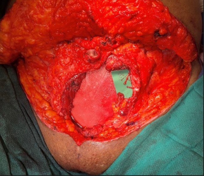
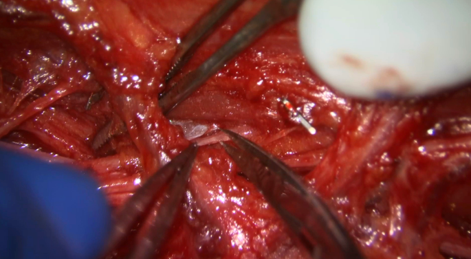
Caroline Leclercq and her team have revolutionized this technique and advocate for “hyperselective neurectomies” [9]. The key differences of their approach was that the neurectomy comprises at least 66% and up to 75% of each branch and it takes place at the most distal entry point of the nerve into the muscle where it starts to terminally branch and arborize. (Figure 1) Additionally, these refinements came at a generation of improved microsurgical skills and techniques [10].
Fig. 1
Distal biceps motor nerve hyperselective neurectomy with transection of 2/3rd of a distal branch with approximately 1/3rd just deep in view remaining to collateralize and maintain muscle strength
Microsurgical Hyperselective Neurectomies by LocationShoulderWhile the pectoralis major, subscapularis, and teres major certainly play some role in the most common deformities of shoulder adduction and internal rotation, accessing their donor nerves for hyper-selective neurectomies is much more difficult. There is significant variability in the pectoralis major motor fibers as well, meaning completely denervating 2/3rd to 3/4ths of each branch is unlikely to be done and recurrences can occur. Simple complete tenotomy of the pectoralis major and lengthening teres major are therefore mainstays of treatment for the shoulder [11].
One exception is the latissimus dorsi, which I consider for treatment second most commonly after the pectoralis major. The motor nerve is singular, consistent, and readily available for hyper-selective neurectomy in a standard supine approach as one would for a latissimus dorsi muscle flap and pedicle dissection. The only data on HSN is on the thoracodorsal nerve and showed long-lasting results with improvement in abduction of the shoulder [6].
Anterior humeral joint instability must be avoided from overzealous anterior approach and lengthening of the subscapularis, and for adults I rarely need to do this at all after treating the pectoralis with a tenotomy, the latissimus with HSN, and teres major with lengthening in that order.
ElbowHSN of the musculocutaneous nerve is a reliable procedure that gives a long-lasting result to the patient. The key is a long incision along about 15–75% of the arm from proximal to distal to ensure identification of the most proximal and most distal branches to the biceps and brachialis, respectively. Technical keys include making this incision just lateral to the bicipital groove so the MABC and basilic vein are kept medial in the medial flap of skin, and the interval between the biceps and brachialis can be readily entered. This also prevents mistaking the median nerve for the musculocutaneous nerve, which is more superficial in the plane of dissection just below brachial fascia under the basilic vein.
The musculocutaneous nerve is found deeper in this interval in the mid upper arm underneath the biceps short and long heads. The motor branches, particularly to the proximal biceps, often run with vascular pedicles that need to be ligated for proper visualization.
It is imperative to understand the variability of the branching patterns and Binder et al. 2014 should be considered required reading [12]. There are typically 1–2 main trunks to the biceps and 2–3 major branches that arise from 1 to 2 main trunks in the brachialis. However, variability is the rule, and a stimulator is key for this dissection. The brachioradialis should be addressed either in tandem or staged depending on factors as discussed above. This is a separate incision between the brachioradialis and the brachialis about 6–7 cm above the lateral epicondyle to just beyond the elbow. There are typically 1–3 small branches to the brachioradialis. Great care must be taken to preserve the ECRL motor branch, which can often run with a main brachioradialis nerve. I have been routinely performing an origin release of the brachioradialis off the humerus to prevent some subtle recurrences in elbow flexion tone along with the HSN.
Outcomes with this technique typically showed that after HSN there was only a transient loss of strength that recovered by 4–6 months follow up without recurrence at up to 7 year follow up [6]. Only incomplete treatments and recurrences noted were when the brachioradialis wasn’t addressed or for large radial nerve branches to the brachialis.
It is important to remember triceps spasticity as well and I perform this concurrently if there is incomplete elbow flexion actively that improves with radial nerve anesthetic blockade. Otherwise, I tend to wait after the first stage if mild and overpowered with the elbow flexor strength. I follow the technique described by Wu et al., where there is triceps z-lengthening with a 2/3rd’s neurectomy of the distal radial nerve branch to the triceps medial head [13].
ForearmBy far the most consistent deformity in the forearm is spasticity and myotendinous contracture of the pronator teres that limits supination. With a curvilinear proximal incision, the interval between the radial artery and superficial sensory branch of the radial nerve uncovers the distal insertion of the pronator teres. The pronator teres is z-lengthened intramuscularly as long as possible. This gives access to the median nerve between the radial artery radially and the flexor-pronator mass ulnarly. The pronator teres branch is typically proximal to the FDS and FCR branches, but there is great variability in these branching patterns that mandate careful stimulation. There are typically 1–2 major nerve trunks (one is more distal) that can undergo hyperselective neurectomy. This same approach gives access to the FDS and FCR branches.
Outcomes data with HSN of the pronator teres shows roughly a 1–2 MAS score reduction in forearm pronator for up to 31 months of follow up [6]. There is evidence of a small recurrence rate of 0.3 T-score in the Tardieu scale between 15 and 31 month follow up, which is more than with elbow spasticity [6].
It is important to note that the pronator quadratus is often another significant portion of the pronator deformity and this nerve is readily available for HSN at the base of the FDP tendons. The failure to address the AIN branches to the pronator quadratus may explain partial recurrence or incomplete treatment in the literature.
Wrist and FingersIf the fingers are fully closed and quick wrist extension elicits a spastic catch, or passive full extension isn’t possible, then treating the FCU and FCR separately is indicated. This is done via the proximal forearm approach as above to Sect. 66%-75% of the main trunk to the FCR. To improve contracture passively, I will also z-lengthen the FCR distally through a separate, distal curvilinear incision that gains access to the extrinsic finger flexors and the distal FCU. The FCR has a short musculotendinous junction that makes fractional lengthening not efficacious [14].
If the patient has volitional wrist flexion I usually just do a tenotomy of the PL or transfer it to the APL or APB for improved radial thumb abduction. If I have poor volitional wrist flexion, and if I am using the FCU or FCR as an EDC tendon transfer, I z-lengthen the PL. It is imperative to keep wrist flexion volitionally intact, if present and separate from finger flexion, to allow tenodesis finger opening.
I typically fractionally lengthen the FCU and do not address the motor nerve with HSN unless there is recurrent wrist spasticity or there is a 90-degree contracture with marked FCU tone. To access the FCU branches, I make a separate incision about 5–6 cm distal to Osbourne’s ligament and find the 2–3 FCU branches on stimulation and HSN each branch. This is also the same surgical approach that one could access the FDP branch from the ulnar nerve, which normally arises 5 cm from the epicondyle to innervate the FDP muscles except for the index finger [14].
Because the extrinsic finger and thumb flexor innervation is variable, requires extensive intramuscular dissection of the FDS to isolate, and typically has elements of spasticity and contracture, improving spasticity is more efficiently done with tendon lengthening [15]. Because the spasticity is indirectly dealt with, however, this approach yields about a 9% recurrence rate in the literature, and this matches my 10% in my practice well [15].
For recurrences and the rare patient that has pure spasticity without spastic contractures, I use HSN FDS motor branches, the FDP motor branches from the median and ulnar nerve, and the AIN branches to FPL and index FDP as described by Emamhadi et al. [16]. There are usually 2–3 FDS branches, the distal of which can arise in the distal forearm, 1 median and 1 ulnar nerve contribution to the FDP, and the FPL and FDP index get 1–2 main trunks from the distal AIN. A technical tip is that when a branch is isolated the distal continuation of the median, ulnar, or AIN is stimulated and if there is no longer finger flexion, then intramuscular dissection doesn’t need to continue further as it is a purely sensory at this point.
Results with hyperselective neurectomies are promising, which mostly show complete resolution of spasticity with mild recurrences of 0.4 on MAS scales up to 31 months [16]. Transient weakness resolved by 6 months.
IntrinsicsIf there are obviously contracted intrinsics on Finochietto testing then I will do Littler releases. This includes cutting the contributions of the interossei and lumbricals to the lateral bands via the oblique fibers while maintaining the transverse fibers of the sagittal band. It is important to do this on both the radial and ulnar side of each finger in spasticity to prevent deviation post-op.
I will do these releases during the first stage of spasticity surgery along with the procedures discussed above from shoulder to intrinsics. [Figure 2] However, if they have pure spasticity without contractures or it is difficult to assess either the intrinsics or thumb in the setting of spastic contractures of the extrinsic finger flexors, then I will address them in the second surgery. This also allows fine tuning tension of tendon transfers or adding additional transfers. At this point separate blocks of the ulnar motor or thenar recurrent can help identify intrinsic spasticity or thumb-in-palm causality.
Fig. 2
(A) Typical incision pattern to perform tenotomy of the pectoralis major, HSN of the musculocutaneous nerve, origin release of the BR and HSN BR motor nerves, pronator teres HSN and z-lengthening, FCR HSN, FCR z-lengthening, FDP, FDS, FPL, FCU fractional lengthening, and recurrent motor HSN with carpal tunnel release. (B) Incision and design for Littler intrinsic releases for spastic contracture and positive Finochietto test
If spasticity without contractures is present in the thumb-in-palm deformity, then HSN of the thenar motor nerve or the adductor pollicis or FDI is possible [17]. If contracted, then an origin release of the FPB or adductor can be done in conjunction [3, 7].
I commonly encounter intrinsic spasticity pronounced in the long, small and ring finger with a relatively volitional key pinch. Historically this was treated as a complete ulnar motor neurectomy or individual interosseous branches in the palm, which is technically difficult. To save volitional pinch, this can be dealt with instead by a “selective” neurectomy of the ulnar nerve just distal to the hook of hamate with an extended Guyon canal approach. The fascicle to the FDI and adductor is always radial and volar to the hypothenar and other interosseous branches. (Figure 3) With microscope dissection one can cut the rest of the ulnar nerve to treat intrinsic spasticity without sacrificing key pinch. My personal series on this is 12 patients without recurrence at 1.5 years that had no change in key pinch strength 6 post-op [18].
Fig. 3
(A) Dissection of the ulnar motor nerve into ADM, hypothenar, and combined adductor/FDI branch + interosseous branches. All branches would be cut but the ADM/FDI to keep volitional pinch while treating intrinsic spasticity. (B) Typical branching pattern and fascicular anatomy of the deep ulnar motor at the hook of hamate
Tendon-Based ProceduresIt is increasingly recognized that function sparing tendon lengthening procedures without addressing the tone with neurectomies can result in higher recurrence rates [15].
Additional tendon procedures, other than the ones described above, are done to rebalance weak muscles. These commonly include ECU to ECRB, PL to EPL, FCR to APL or APL tenodesis, FDS opponensplasty, and anti-clawing procedures (I prefer the Stiles- Bunnell with lengthened FDS).
Joint/Bone ProceduresI remain very cautious of joint arthrodesis except for the most severe cases of wrist flexion contracture with carpal pathology and for thumbs with diseased and contracted CMCJs. Even subtle joint range of motion, particularly at the wrist, can provide tenodesis effects for digital opening which can be imperative to maintain in the weakened patient. A MCPJ arthrodesis is my most common bone procedure for spasticity and is for older patients with hyperlaxity in ulnar deviation (usually iatrogenic from therapy with a spastic adductor or FPB). Keep in mind these patients often have disuse osteopenia with thin overlying skin and plates can be problematic.
Post-Operative ProtocolsFor the common flexion deformity at the wrist/elbow/fingers and thumb, I typically splint patients with a forearm-based wrist neutral extension with thumb abducted for 3–4 days before starting an early active range of motion protocol. If the pronation deformity has been addressed with a tendon procedure, then I use a muenster splint with thumb abduction. This is done during the day with frequent removal (5-6x daily) for stretching and range of motion for 4 weeks, and then at night for 2–4 more weeks depending on the severity of contractures. If a pure HSN case is performed, I do not splint after the first week and switch to compression garments/sleeves.
Comments (0)