
Remember me


A total of 325 male and 325 female volunteers between 12 and 24 years of German nationality were enrolled between 2021 and 2023 via advertisements in newspapers, flyers and social media. The groups were each composed of 25 participants per year of age and sex and filled consecutively determined by the applicants’ submission date. Since the study aimed at examining the physis’ development in healthy individuals, the inclusion criteria were only composed of known sex and proven age. Further noted characteristics of the volunteers were known illnesses as well as past and present medication. The exclusion criteria were developmental diseases and disorders, injuries to the knee joint area, in order to prevent the inclusion of individuals with hitherto unknown adverse post-traumatic changes to the physiological skeletal maturation, and common contra-indications of low-field MRI, especially incorporated metal elements and potential gravidity. Noteworthy, due to the construction of the scanner claustrophobia was no exclusion criterion. This study received approval by the responsible ethics committee and a signed written informed consent from each volunteer or in case of minors from the legal guardian was obtained. The study’s setting is prospective and the examined volunteers’ data was used to establish the dataset for the project “age estimation in refugees of questionable minority” that was funded by the European Asylum, Migration and integration Fund (AMIF) and the German Federal Office for Migration and Refugees.
Scanner and sequencesThe MRI-scans were performed with a 0.31 T scanner (Esaote O-Scan Premium, Esaote S.p.A.) using a dedicated dual phased array (DPA) coil for the wrist. A proton density-weighted (PDw) spin echo (SE) sequence in Dixon technique (Dixon, sequence trade name SPED PD), in coronal orientation was acquired (TR 1800 ms; TE 20 ms / 31 ms; duration 6:16 min; slice thickness 3.5 mm; measured matrix 288 × 288; measured pixel size 0,56 × 0,56 mm²; reconstructed matrix 512 × 512; reconstructed pixel size 0,31 × 0,31 mm²) from which fat-sensitive water-suppressed (fat-only) and water-sensitive fat-suppressed (water-only) series were computed.
For the morphological assessment of the stages the images were viewed on a PACS-workstation with simple post processing allowed i.e. window leveling. Firstly, all cases were evaluated by an examiner with ten years of experience in musculoskeletal MRI diagnostics and age estimation (C.O.) and in order to determine the intraobserver-agreement a re-evaluation of all randomly rearranged cases was performed after a lapse of more than 6 months so as to prevent recall-bias. Secondly, an examiner with more than 20 years of experience in musculoskeletal MRI diagnostics and age estimation (V.V.) independently evaluated all cases for determining the interobserver-agreement. The evaluations were performed without knowledge of sex, age and earlier evaluations of the examined volunteers. Lastly, the assessed stages were then tested against the proven age.
Low-field classificationThe study group decided to use a PDw Dixon sequence. The rationale behind this decision was, that the Dixon technique could provide morphological information about osseous structures via the fat-only series and about watery structures via the water-only series, all in a single scan and with elevated sensitivity towards watery signals. After reviewing the acquired data, it became obvious that relevant landmarks of the distal radius’ physis were identifiable and therefrom a classification was construed that builds upon the synergistic use of the acquired images. The fat-only series is used to evaluate the osseous characteristics of the epiphysis and locate the physis or in later phases the physeal scar. Then, the water-only series is used for the purpose of detecting watery components within the examined osseous structures in the continuity of the physis. Thusly, it is possible to assess a stage by certain changes of the physeal ossification that are associated with the individual’s age. While stages 1–5 can already be differentiated solely in the fat-only sequence, the appearance of stages 5 and 6 are virtually identical in the fat-only sequence. In these stages, the water-only sequence is used to look for a hyperintense signal in the continuity of the epiphyseal scar. In stage 5, this hyperintense signal is present, while in stage 6 no such hyperintense signal can be found, marking the end of discernable morphological changes of the fusion as recognized by this classification. Furthermore, as the cohort’s lower bounds lay at 12 years of age, the acquired images can’t have captured the earliest developmental features of the physis. To signal awareness of this fact stage 1 was therefore left blank and the classification starts with stage 2.
Corresponding schematics were produced for the distal radius’ physis accompanied by examples of the stages (see Figs. 1 and 2).
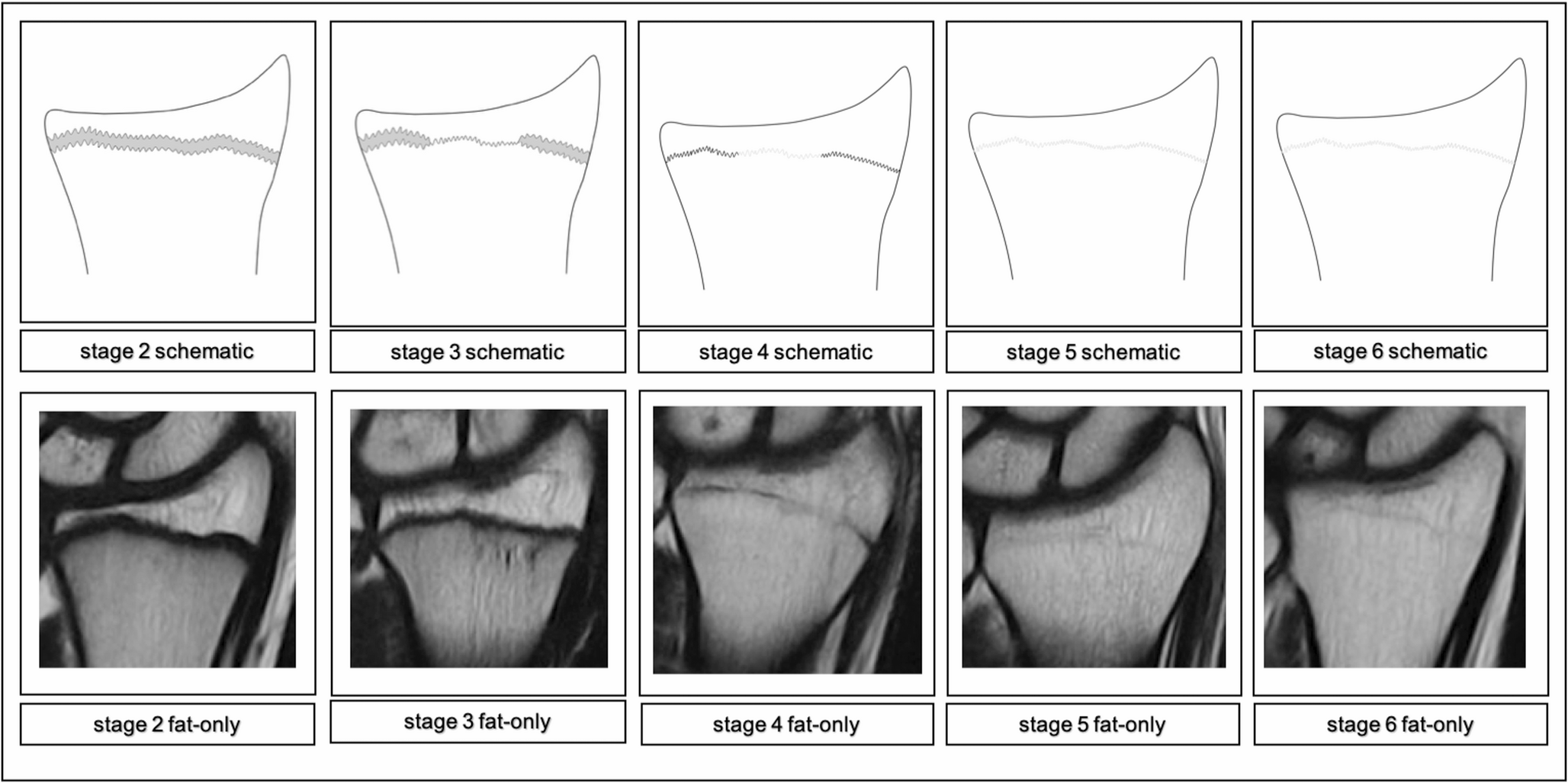
Fig. 1
Schematic drawings for the stages in the PDw Dixon sequence fat-only series and examples (0.31T; non-contrast enhanced; coronal slice orientation); from left to right: male 14yrs, male 15yrs, male 17yrs, male 22 yrs, female 24yrs
Fig. 2
Schematic drawings for the stages in the PDw Dixon sequence water-only series and examples (0.31T; non-contrast enhanced; coronal slice orientation); from left to right: male 14yrs, male 15yrs, male 17yrs, male 22 yrs, female 24yrs
Following is a comprehensive description of the stages’ key characteristics in the fat-only series and the water-only series adapted to low-field imaging:
Stage 2 In the fat-only series a continuous, broad band of low signal intensity is discernable. The band’s borders are smooth or serrated and towards the diaphysis the border may be blurry.
The water-only series shows a thick, hyperintense line congruent to the band of the corresponding fat-only series with smooth or serrated borders. Towards the diaphysis the border may be blurry, forming a band. In the course of the band layers of high signal intensity may be discernable, that can be continuous or discontinuous and sporadically convening.
Stage 3 In the fat-only series a discontinuous, broad band of low signal intensity is discernable. The band’s borders are serrated towards the epiphysis and the diaphysis. The band sporadically narrows, forming a single, thick serrated line of low signal intensity.
The water-only series shows a thick line of high signal intensity in the course of the band of the corresponding fat-only series, that is segmentally tapering into a continuous, thin serrated line of high signal intensity.
Stage 4 In the fat-only series a discontinuous, thick serrated line of low signal intensity between the epiphysis and the diaphysis is discernable with thinner segments of intermediate signal intensity.
The water-only series shows a continuous, discontinuous or dotted, thin line of hyperintense signal in the continuity of the line of the corresponding fat-only series.
Stage 5 In the fat-only series a continuous, thin serrated line of intermediate signal intensity between the epiphysis and the diaphysis is discernable.
The water-only series shows a discontinuous or dotted, thin serrated line or spots of hyperintense signal in the course of the thin line of the corresponding fat-only series.
Stage 6 In the fat-only series a continuous, thin serrated line of intermediate signal intensity between the epiphysis and the diaphysis is discernable. The water-only series shows no hyperintense signal in the course of the thin line of the corresponding fat-only series.
Statistical analysesStatistical analyses were conducted using IBM SPSS Statistics 28 for Mac OS X (release 25th May, 2021, Build 28.0.1.1, IBM Corporation). Minimum, maximum, mean ± standard deviation and median with lower and upper quartiles were defined for each stage of the classification to find the minimal ages. Intra- and interobserver-agreements were determined by calculating the kappa coefficients. Sex related differences in the stage-assessment across the ages were analyzed using the Mann-Whitney-U Test to determine their statistical relevance (p < 0.05, exact, two-tailed).
Comments (0)