
Remember me

The solubility of free DCB in water was evaluated under various temperature and time conditions while protected from light (Fig. 2(a)). At room temperature, the solubility of DCB was determined to be 1.232 ± 0.043 mg/mL at 3 h, which decreased to 1.036 ± 0.026 mg/mL at 12 h, representing a 15.9% reduction. This decrease in solubility over time is likely attributed to hydrolytic degradation of DCB, suggesting that prolonged exposure to aqueous environments at room temperature leads to reduced chemical stability. In contrast, at 37 °C, the solubility of DCB was observed to be 1.948 ± 0.031 mg/mL at 3 h and slightly decreased to 1.915 ± 0.025 mg/mL at 12 h. Although this difference was not statistically significant, the downwards trend in solubility still suggests the occurrence of hydrolytic degradation at elevated temperatures. The relatively higher solubility observed at 37 °C than at room temperature highlights the influence of temperature on enhancing the solubility of DCB in water. Overall, these findings underscore that while temperature can increase DCB solubility, time-dependent degradation due to hydrolysis remains a concern, particularly under prolonged aqueous exposure.
Fig. 2
a Solubility profile of free DCB in water at room temperature and 37 °C over time (mean ± S.D., n = 3). Stability of free DCB at different temperatures in b water and c 2% citric acid aqueous solution (mean ± S.D., n = 3)
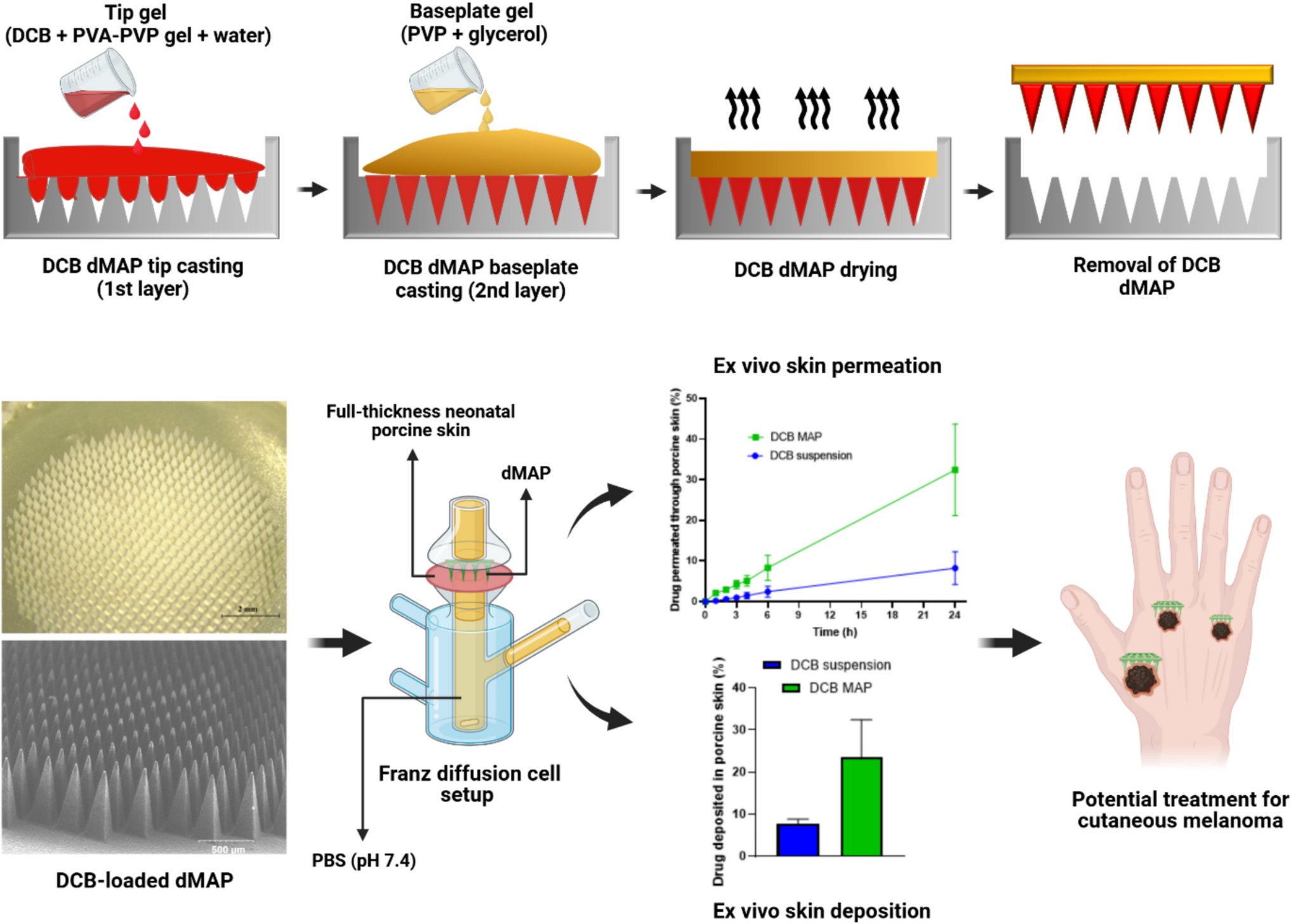
The fabrication of DCB-loaded bilayer dMAP involves multiple steps (42, 53), which expose the DCB to an aqueous environment (PVA-PVP gel) for several hours (24–48 h) until the final dried bilayer dMAP is obtained. Solubility studies have shown that DCB degrades in aqueous environments, even when protected from light. Therefore, to accurately evaluate the hydrolytic degradation of free DCB under various temperature conditions, a controlled study was conducted by introducing 5 µg/mL DCB into aqueous media (Fig. 2(b, c)). Stability assessments were performed at multiple time intervals and temperature settings, with protection from light. Additionally, the potential stabilizing effect of 2% citric acid aqueous solution was investigated, given its known role in enhancing DCB solubility and its inclusion in FDA-approved DCB powder for injection formulations. The primary objective of this study was to elucidate the effects of temperature and time on the hydrolytic degradation of DCB. At 24 h, DCB in water retained 98.41 ± 3.01% of its initial concentration at room temperature, whereas only 80.13 ± 0.72% remained at 37 °C, reflecting a 12.49-fold increase in degradation at elevated temperatures. After 168 h (7 days), the stability gap widened further, with 92.52 ± 1.66% of the DCB preserved at room temperature, whereas only 39.93 ± 1.87% was preserved at 37 °C (p < 0.05). Interestingly, compared with water alone, the presence of 2% citric acid (aqueous) did not significantly alter the degradation profile of DCB, suggesting that while citric acid improves solubility, it does not confer additional stability against hydrolytic degradation in aqueous environments (p > 0.05). These findings highlight the crucial need to control the temperature and protection from light when free DCB is used, particularly during the fabrication of bilayer dMAP. In typical micromoulding processes, bilayer dMAP is dried at 37 °C for at least 12 h to remove moisture and prevent the needles from deforming when applied to the skin (48). However, this drying step can accelerate the degradation of DCB. Considering the results of the stability study, an alternative drying method was adopted to remove moisture from the bilayer dMAP without exposing DCB to high temperatures, which helped reduce degradation and maintain the stability of the compounds.
Fabrication and Morphological Assessment of DCB-loaded Bilayer dMAPThe DCB-loaded bilayer dMAP was successfully fabricated, incorporating 600 pyramid-shaped needles uniformly arranged within a 0.75 cm2 circular area. Each needle had a base width of 300 µm, a height ranging from 700 to 750 µm, and was spaced 50 µm apart from adjacent needles. PVA and PVP were used to fabricate bilayer dMAP tips. The primary reasons are their safety, skin compatibility, mechanical properties, and excellent dissolution behaviour. While PVA provides flexibility and PVP contributes rigidity, their combination results in a more robust needle structure, likely due to hydrogen bonding between their functional groups (54, 55). Moreover, because the base of the bilayer dMAP tips may not fully penetrate the skin, drug delivery from the baseplate can be inefficient. Thus, DCB was specifically loaded into the tip region, while the baseplate was composed of a drug-free PVP and glycerol gel, reducing the likelihood of drug loss (44).
To develop an optimized DCB-loaded bilayer dMAP, five different formulations of drug-loaded tip-casting gels were evaluated, as summarized in Table 1. The structural integrity and drug distribution within the bilayer dMAPs were assessed via a Leica EZ4D digital light microscope to confirm successful needle formation and uniform drug localization at the tips. Formulation F1 failed to form needle tips because of the high viscosity of the drug‒polymer gel in the absence of added water, preventing proper mould casting. In contrast, formulations F2 to F5 successfully produced bilayer dMAPs. While F2 could be cast onto the mould, its viscosity remained sufficiently high to cause premature drying before the gel could fill all microcavities after centrifugation, resulting in incomplete MAP tip formation, as shown in Fig. 3(a, e). Formulations F4 and F5 achieved complete MAP tip formation without structural breakage. However, the increased water content in these formulations led to insufficient homogeneity in the drug‒polymer mixture. Consequently, the resulting MAP tips exhibited uneven drug distributions, as illustrated in Fig. 3(c, d, g, h). Among all formulations, F3 yielded bilayer dMAP with well-formed, intact tips and uniform drug distribution throughout the tips, as shown in Fig. 3(b, f). This highlights the importance of optimizing the water content in the tip-casting gel to achieve both structural integrity and consistent drug loading. Further confirmation through SEM revealed that bilayer dMAP fabricated using F3 possessed sharp, undeformed needle structures with densely packed drug contents in the tips, as illustrated in Fig. 3(i, j).
Fig. 3
Morphology of DCB-loaded bilayer dMAPs. For each formulation, the same physical array is shown from two viewpoints; top-down a–d and oblique/slant (~45°) e–h: F2 a, e, F3 b, f, F4 c, g, F5 d, h. All panels a–h were captured on intact arrays immediately after demoulding/drying and before any compression or insertion experiments; no “after” images are included in this figure. Panels i and j are SEM micrographs of a representative array made with the optimised formulation F3: i array overview and j higher magnification with tilted view. (Each pair, e.g., a↔e, shows the same array)
Mechanical Properties and Insertion Characteristics of DCB-loaded Bilayer dMAPThe bilayer dMAP is required to exhibit adequate mechanical strength to ensure reliable penetration of the skin without structural failure, thereby enabling effective dermal drug delivery (56). To evaluate this property, mechanical and insertion tests were performed on the DCB-loaded bilayer dMAP. According to previous studies, a force of approximately 32 N represents the maximum force that can be applied by a human for MAP insertion into skin tissue (44). In the present study, mechanical strength was assessed by quantifying the percentage reduction in needle height following the application of a 32 N force per patch for 30 s against a metal block. As depicted in Fig. 4(a, b), the DCB-loaded bilayer dMAP demonstrated a needle height reduction of 6.16 ± 0.13%. To further assess the mechanical strength of the bilayer dMAP under simulated conditions, an additional test was performed using layered parafilm as a model for the skin. The same force of 32 N was applied, and the resulting reduction in needle height was only 3.18 ± 0.64% (Fig. 4(c, d)). This minimal deformation indicates that the DCB-loaded bilayer dMAP maintain their structural integrity under insertion-like conditions, reinforcing their potential for effective dermal drug delivery (Fig. 4(e)).
Fig. 4
Mechanical strength, insertion characteristics and drug content of DCB-loaded bilayer dMAP . Digital microscopy images showing the same bilayer dMAP arrays before and after compression at 32 N/array for 30 s using a TA-XT2 texture analyser: a before and b after compression against a metal block; c before and d after compression against eight layers of Parafilm® M. e Percentage reduction in needle height post-compression (mean ± SD, n = 3). f Insertion profile of the bilayer dMAP (mean ± SD, n = 3). g–j Microscopy images of individual Parafilm® M layers from the insertion study: g first, h second, i third, and j fourth layers. k Drug content in the tips and baseplate of the bilayer dMAP (mean ± SD, n = 3)
The insertion capability of the DCB-loaded bilayer dMAP was examined via a Parafilm® M-based skin simulant model, which consists of eight stacked layers, each approximately 1 mm thick (53). This model approximates the average human skin thickness of 1.19 mm, serving as a practical surrogate for evaluating MAP penetration. A force of 32 N per patch was applied for 30 s, and the extent of penetration was determined by calculating the proportion of punctures in each layer. As presented in Fig. 4(f-j), the bilayer dMAP successfully pierced 100 ± 0% of the first Parafilm® M layer, 92.16 ± 6.6% of the second layer, and 3.33 ± 1.42% of the third layer, indicating an estimated penetration depth of approximately 390 µm. Furthermore, our previously published works have confirmed the successful insertion of MAPs into excised neonatal porcine skin using the optical coherence tomography technique (37). Considering that the stratum corneum measures approximately 20 µm (57) and that the epidermis extends between 75 and 270 µm (58), the achieved depth suggests that the MNs can effectively reach the upper dermis. These regions are critical for targeting early-stage cutaneous melanoma (up to 2 mm), confirming the suitability of bilayer dMAP for dermal drug delivery.
Determination of the DCB Content in Bilayer dMAP Tips and BaseplatesThe tips of the DCB-loaded bilayer dMAP were fabricated via a formulation composed of DCB, PVA-PVP gel, and water, resulting in a drug loading of 66.66% within the dMAP tips. Notably, the remaining 33.34% polymer content was sufficient to impart adequate mechanical strength to the dMAP tips, enabling them to withstand a 32 N force without significant needle deformation. Quantitative analysis via UV‒Vis spectrophotometry (Cary 60, Agilent Technologies Ltd., UK) at a wavelength of 330 nm revealed that DCB-loaded bilayer dMAP tips contained 4.33 ± 0.52 mg of DCB, whereas the base plate contained 0.68 ± 0.16 mg, amounting to a total of 5.01 ± 0.69 mg of DCB per patch (Fig. 4(k)). Although DCB was initially loaded exclusively into the MAP tips, a minor level of back-migration to the baseplate was observed during the drying phase, likely due to the hydrophilicity of DCB. This phenomenon has also been reported in previous studies (39, 59,60,61,62). While some studies have reported substantial back migration of hydrophilic drugs from the tip region to the base plate, the current study revealed minimal back migration of DCB (~ 15%). Nevertheless, most of the DCB remained localized within the MAP tips, indicating successful containment and minimal diffusion-related loss during fabrication.
Dissolution Behaviour of DCB-loaded Bilayer dMAPThe dissolution properties of bilayer dMAP are essential for ensuring successful drug release into the skin (43). In this investigation, their dissolution was assessed via the use of excised full-thickness neonatal porcine skin as a model. The DCB-loaded bilayer dMAP was inserted into the tissue using thumb pressure for 30 s and then maintained at 37 °C. At specific intervals, the patch was gently removed and analysed with a Leica EZ4D digital light microscope to evaluate the progression of bilayer dMAP tip dissolution. Figure 5(a-d) shows the dissolution profiles of the DCB-loaded bilayer dMAP. Within the first 5 min of insertion, slight dissolution of the bilayer dMAP tips was observed. By 30 min, a substantial portion of the tips had dissolved, and complete dissolution of the entire bilayer dMAP tip was achieved within 60 min. The dissolution performance of bilayer dMAP is influenced by several factors, including skin moisture content, drug solubility, drug loading percentage in the bilayer dMAP tips, polymer concentration, needle density within the patch, and the insertion efficiency of the bilayer dMAP. Given the interplay of these variables, achieving full dissolution within 1 h is considered satisfactory. Interestingly, despite the observed insertion depth being limited to approximately 390 µm, all the bilayer dMAP tips (measuring 700–750 µm in height) were found to have fully dissolved. This suggests a sequential dissolution mechanism: as the portion of the bilayer dMAP tips embedded in the skin begins to dissolve, it allows the remaining length to progressively come into contact with skin moisture, thereby facilitating continued dissolution. This behaviour may also explain the longer duration (up to 1 h) required for the complete dissolution of the dMAP tips. Nevertheless, upon application of the bilayer dMAP for 15 min, DCB was observed to accumulate within the excised porcine skin, forming localized drug depots, as illustrated in Fig. 5(e, f). The formation of these depots suggests that the bilayer dMAP tips effectively penetrated the stratum corneum and delivered the drug into deeper layers of the skin. Such intradermal depot formation is beneficial, as it facilitates sustained drug release at the site of administration, potentially enhancing therapeutic efficacy in cutaneous melanoma treatment while minimizing the risk of immediate systemic exposure.
Fig. 5
Digital microscopy images showing the dissolution of DCB-loaded bilayer dMAPs upon insertion into full-thickness neonatal porcine skin at a 5 min, b 30 min, c 45 min, and d 60 min. Images e and f depict drug depot formation in the skin following a 15-min application of bilayer dMAP
Ex vivo Pore Visualization StudiesAlthough our previous study indicated that a 15-min application of bilayer dMAP delivers DCB into the deeper layers of the skin, it remains uncertain whether true intradermal depot formation occurred or if the drug merely resided on the surface of the skin. This ambiguity arose from the imaging technique, which involved capturing microscopy images from the top view of DCB-deposited excised porcine skin. To confirm actual skin penetration, a pore visualization study was performed via methylene blue staining. Following manual insertion of the DCB-loaded bilayer dMAP into porcine skin for 30 s via thumb pressure, 200 µL of methylene blue solution (1 mg/mL) was applied to the skin surface for 15 min. The dye was then removed, and the skin was examined via a Leica EZ4D digital light microscope. Figure 6(a-i) shows the formation of small pores in porcine skin following the application of bilayer dMAP. Interestingly, despite the bilayer dMAP tips having a base width of 300 µm, the observed pores on the skin were relatively small. This can be attributed primarily to the limited insertion depth of the bilayer dMAP, which achieved only 52% needle length penetration into the Parafilm® M model, corresponding to approximately 390 µm. Furthermore, the inherent elasticity of the skin likely caused the pores to shrink immediately after bilayer dMAP tip withdrawal.
Fig. 6
a-e Steps involved in the ex vivo pore visualization study using methylene blue on full-thickness neonatal porcine skin. Digital microscopy images of f untreated skin (16 ×) and methylene blue-stained skin at g 16 ×, h 25 ×, and i 35 × magnifications. Ex vivo profiles of j permeation and k deposition of DCB from bilayer dMAP and suspension using full-thickness neonatal porcine skin (mean ± S.D., n = 3). (l) Drug content in bilayer dMAP on days 0 and 8 of storage at room temperature protected from light (mean ± S.D., n = 3)
Ex vivo Permeation and Deposition StudiesThe ex vivo permeation and deposition profiles of DCB from the dMAP and suspension formulations were evaluated via full-thickness neonatal porcine skin in a Franz diffusion cell apparatus (42, 56). The two delivery systems were compared to assess their relative efficiency in dermal drug delivery. During the first hour, the DCB-loaded bilayer dMAP permeated 2.1 ± 0.38% of the drug across the skin, which was significantly greater than the 0.2 ± 0.1% observed with the DCB suspension. By the end of 24 h, the cumulative permeation from the bilayer dMAP reached 32.43 ± 11.2% (1.62 ± 0.56 mg/0.75 cm2), whereas the suspension reached only 8.24 ± 4.03% (0.41 ± 0.2 mg/0.75 cm2), representing a 3.93-fold increase in drug permeation from the bilayer dMAP (Fig. 6(j)). The limited permeation observed with the DCB suspension can be attributed to the hydrophilic nature of DCB, which has a measured solubility of 1.232 ± 0.043 mg/mL at 3 h. Although this study primarily aims to retain DCB locally within the skin, some drug permeation was observed. This is not intended for systemic delivery but reflects the drug’s local diffusivity. The permeated drug likely interacts with melanoma cells near the application site before any systemic absorption, thereby still contributing to local therapeutic efficacy. Although systemic exposure to DCB via MAPs may cause some rare or less common side effects, the primary side effects associated with the current clinical method of intravenous administration, such as redness, pain, or swelling at the injection site, can largely be avoided by using MAPs. However, this should be further confirmed through in vivo studies using a suitable animal model (63).
In addition to permeation, drug deposition within the skin was also assessed at 24 h. The bilayer dMAP group presented significantly greater drug retention, with 23.52 ± 8.87% (1.17 ± 0.44 mg/0.75 cm2) of DCB deposited in the skin than the 7.77 ± 1% (0.38 ± 0.05 mg per 0.75 cm2) of DCB in the suspension group. In total, the DCB-loaded bilayer dMAP delivered 55.95 ± 14.47% (2.79 ± 0.5 mg/0.75 cm2) of the drug through both permeation and deposition, whereas the suspension formulation delivered only 16.01 ± 4.53% (0.79 ± 0.12 mg/0.75 cm2) (p < 0.05) (Fig. 6(k)). Notably, stability studies of free DCB (Section Solubility and stability of free DCB) indicate that approximately 20% of the drug was lost due to hydrolytic degradation at 37 °C over 24 h, suggesting that a portion of the DCB was degraded in both the suspension and bilayer dMAP formulations during ex vivo permeation and deposition studies. Despite the occurrence of degradation, a portion of the drug in both the suspension and bilayer dMAP formulations remained neither permeated nor deposited. In the suspension group, this unabsorbed fraction was likely due to the limited ability of the hydrophilic DCB to effectively penetrate the stratum corneum. In the case of bilayer dMAP, the nondelivered drug is primarily attributed to incomplete MAP insertion (52%), as demonstrated in earlier insertion studies (Section Mechanical properties and insertion characteristics of DCB-loaded bilayer dMAP), where the full length of the needle did not completely penetrate the skin. Collectively, the results of the permeation and deposition studies clearly demonstrate the superior performance of the DCB-loaded bilayer dMAP compared with that of the conventional topical delivery system (suspension). The enhanced ability of the bilayer dMAP to deliver a significantly higher amount of drug to the target site supports its potential as an effective strategy for the localized treatment of cutaneous melanoma.
Short-term Storage Stability of DCB-loaded Bilayer dMAPDCB is highly sensitive to environmental factors such as light, temperature, and moisture (23,24,25,26,27,28). As shown in the stability studies of free DCB (Section Solubility and stability of free DCB), approximately 7.5% of the drug degraded over a 7-day period under room temperature aqueous conditions, even when protected from light. Gel-based formulations are widely used for topical drug delivery because of their simplicity of application. However, their stability becomes a concern when moisture-sensitive compounds such as DCB is loaded. This challenge is particularly relevant given the importance of formulations that remain stable at room temperature without requiring refrigeration, a key consideration for enhancing global access to medications and improving the efficiency of drug distribution. Considering these limitations, the current study evaluated the short-term storage stability of DCB-loaded bilayer dMAP over an 8-day period at room temperature, with protection from light. After 8 days, the DCB content was quantified, revealing 4.1 ± 0.65 mg of DCB in the MAP tips and 0.95 ± 0.2 mg in the baseplate, totalling 5.05 ± 0.86 mg per dMAP (p > 0.05). These results indicate that no measurable degradation of DCB occurred during the storage period (Fig. 6(l)). This stability is attributed to the dry state of the formulation, which effectively prevents hydrolytic degradation of DCB in the absence of moisture. Given these findings, bilayer dMAP is a more robust and stable alternative to conventional topical formulations for delivering DCB in the treatment of cutaneous melanoma. Moreover, unlike gels that expose the drug directly to light upon application, the bilayer dMAP design could offer an added layer of protection. By incorporating the drug exclusively in the MAP tips and using a drug-free baseplate that can be covered with a light-shielding backing layer, the bilayer dMAP minimizes light-induced degradation and enhances drug preservation during delivery.
Comments (0)