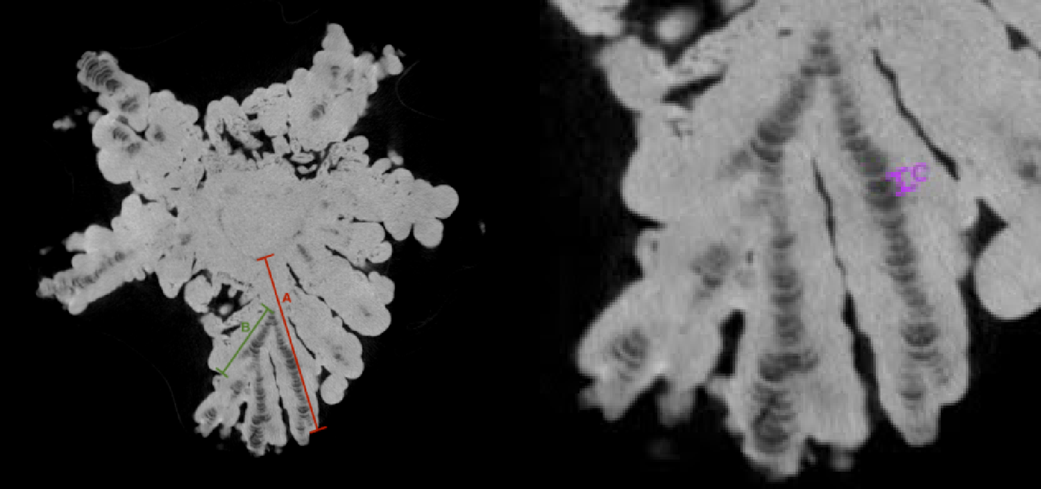
This study demonstrates a new micro-CT–based morphometric method for quantifying layering patterns in jackstone calculi, providing a reproducible framework for evaluating jackstone growth mechanisms. Using this method, we show that the jackstone arms exhibit more numerous and thinner layers toward their distal tips, supporting a model of abrasion-mediated remodeling during stone growth.
In addition to the microstructural findings, the anatomical context of the donor provides important physiological insight into jackstone formation. The markedly enlarged prostate and pronounced bladder wall hypertrophy are anatomical changes commonly associated with chronic bladder outlet obstruction and urinary stasis, a well-established risk factor for urinary stone formation [26,27,28]. For reference, the average bladder weight in adult males is approximately 25–30 g [29], whereas the donor’s bladder weighed 73.2 g. Similarly, the donor’s prostate measured 90.7 g, substantially exceeding the typical weight of 20–30 g in normal prostates and 30–50 g in benign prostatic hyperplasia [30].
Chronic obstruction increases post-void residual volume and reduces urinary flow, conditions that favor crystal nucleation, retention, and incremental stone growth [26]. As the bladder compensates, muscular hypertrophy and inflammation develop, contributing further to urinary stasis. In this donor, the combination of bladder wall hypertrophy and significant prostate enlargement supports the interpretation that persistent urinary retention created a biochemical and mechanical environment conducive to jackstone formation [27, 28].
From this clinical context, we then turn to the microstructural patterning revealed by micro-CT imaging, which allows us to evaluate the mechanism by which the arms remodel and extend over time. Our method provides a means by which microstructural patterns may be evaluated and further provides anecdotal support for the hypothesis that the distinctive morphology of jackstone calculi results from the accelerated radial growth of their arms compared to the base, due to physical abrasion between the stone and the bladder wall [4]. Specifically, the distal regions of the arms contact the bladder wall, leading to the stripping of proteins and/or minerals and subsequent exposure of the arm tips. This exposure likely increases remodeling at these sites, promoting further growth, and as the jackstone arms grow, abrasion becomes more frequent, leading to a redundant cycle. The repetitive nature of this abrasion and binding cycle results in the formation of progressively thinner concentric layers over time, as supported in our findings.
Our method provides a technique that would allow the abrasion-driven growth model to be tested, elucidating the unique morphology of jackstone calculi. Canela et al. (2022) suggested that post-abrasion, the exposed protein layer attracts additional proteins, thereby extending the protein layer [4]. If this were the case, an increase in layer thickness, particularly of the radiolucent protein layer, would be anticipated. However, our observations do not corroborate this, as we did not detect evidence of protein-to-protein increased attraction leading to thicker protein layers. Instead, our results indicate that while abrasion may facilitate preferential binding, it does not necessarily result in increased protein layer thickness. This suggests a modified understanding of the growth mechanism, where abrasion influences the pattern of layer remodeling without significantly altering protein layer thickness. These insights contribute to a more nuanced comprehension of the formation and development of jackstone calculi.
This study enhances current knowledge of bladder stone formation mechanisms and, with future research building upon it, may help guide screenings, diagnostics, and clinical treatments [31]. With insight into the link between urinary stasis and stone formation, clinicians can implement earlier interventions in high-risk patients with urinary retention. Additionally, a potential therapeutic approach could focus on reducing the abrasion a bladder stone has with the wall to minimize the remodeling and growth.
There were a few inherent limitations to this work. This study examined a single specimen, limiting generalizability, however the purpose of this work was to establish a methodological framework for future sample-comparative studies. The relative rarity of the condition makes the small sample size nearly unavoidable. Another limitation is the postmortem changes that may have occurred in the donor, including tissue breakdown and loss of muscle tone. However, while the organ weights were taken from a donor patient, one study compared the bladder weights of 10 male donor cadavers, with a mean age of 67 ± 21 years, to ultrasound-estimated bladder weights in living patients and found no significant difference between the bladder weights of cadaveric patients and those of the living cohort [29].
Given the apparent association between urinary stasis and jackstone formation, future studies should explore the potential microbial contributions to this process, as culturing and sequencing jackstone specimens may uncover distinct microbial signatures or biofilm communities associated with their development. With the urinary microbiome now recognized as an integral component of urologic health [32], it is plausible that prolonged urinary stasis may foster colonization by specific bacterial taxa that promote or modulate jackstone formation. This hypothesis is supported by emerging evidence linking particular microbial species to the formation of other stone subtypes [33], warranting further investigation into the microbiological underpinnings of jackstone pathogenesis.


Comments (0)